A Glimpse into the Microcosm: Hassanain Qambari's Visionary Triumph in Nikon Small World Competition
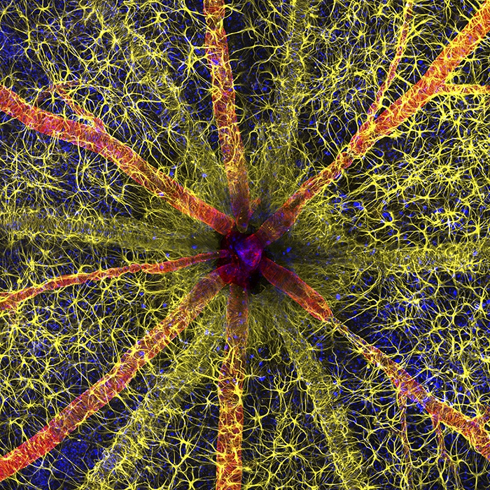
In the mesmerizing realm of microscopic photography, where art meets science, the Nikon Small World Photomicrography Competition stands as a testament to the beauty hidden within the minute. This prestigious global contest has recently announced its 49th annual winners, bringing to the forefront a stunning composition that not only captivates the eye but also contributes profoundly to medical science. Leading the pack with the first-place prize is Hassanain Qambari, alongside collaborator Jayden Dickson of the Lions Eye Institute. Their compelling image of a rodent optic nerve head, vividly displaying astrocytes in yellow, contractile proteins in red, and the retinal vasculature in green, is more than just a picture. It is a critical leap in understanding and combating diabetic retinopathy, a debilitating condition affecting millions worldwide.
As we delve into an exclusive interview with Qambari, we explore the inspiration, challenges, and technical mastery behind this extraordinary image. From his educational journey to the intricate details of capturing the microscopic marvels of the eye, Qambari sheds light on the importance of early detection and innovative research in reversing diabetic retinopathy. His dedication and pioneering work not only earned him the top spot in the Nikon Small World Competition but also promised to inspire a new generation of scientists and enthusiasts in the pursuit of discovery and understanding. Join us as we uncover the story behind the image that has illuminated the complex beauty of the microcosm and heralded a significant stride in medical science.
Educational Journey:
Could you share with us your educational journey and the key experiences that led you to specialize in retinal disease research?
At the finishing of high school, I pursued a degree in Neuroscience and Anatomy and Human Biology at the University of Western Australia before completing a master’s degree with a neuroscience specialization. It was during my dissertation which involved the detection of retinal microvascular changes using a deep learning model in diabetic retinopathy with the Commonwealth Scientific and Industrial Research Organisation (CSIRO). It was from this passion and my understanding of these vascular changes that I progressed onto a PhD through The University of Western Australia at the Lions Eye Institute. I am currently completing my PhD candidature with the Physiology and Pharmacology team at the Lions Eye Institute supervised by the department head, Professor Dao-Yi Yu and co-supervised by Associate Professor. Paula Yu and Clinical Professor. Chandra Balaratnasingam. It is here that I joined part of the team investigating these early vascular changes within diabetic eyes via the novel method of isolated whole eye preparations.
Training and Techniques:
What specific training or techniques have you mastered during your time at the Lions Eye Institute that were crucial in capturing your award-winning image?
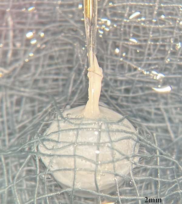
The most important technique I learnt was taking into account all the variables that were crucial in producing a successful stain. These variables include the chemicals used, the concentration used and the duration of exposure (over exposure to certain chemicals like triton x-100 which helps with the penetration of the cellular markers means damage to the cellular structures or in extreme circumstances completely dissolving the tissue). However, before even getting to this stage of trialling different protocols, I first had to develop the fine motor skills for the isolated perfusion eye preparation. This technique requires the dissection of a single vessel called the Ophthalmic artery, approximately 100 microns in diameter and a glass pipette placed inside. Through a process of trial and error I was able to develop the skill of perfusing histological perfusion staining.
One more thing to note is, this image was produced through the combination of perfusion and float staining. Float staining is a histological technique involving the immersion of the tissue in the necessary solutions. This required further trial and error as the concentration and duration of chemical exposure was important on whether the structures stained for via perfusion were damaged or the tissue itself dissolved.
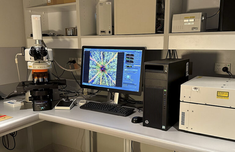
Scanning of the specimen with the confocal microscope was probably the least troublesome, as any error made during the image capturing process could be re-attempted immediately and there are far less variables to consider.
Motivation for the Competition:
What inspired you to participate in the Nikon Small World Photomicrography Competition, and what message did you hope to convey through your submission?
Last year I was troubleshooting some issues regarding my confocal imaging and that’s where I stumbled across the Nikon Small World Competition. Jayden and I both went through the past years’ winners, and we thought it would be a good idea to submit an entry as some of the images we were capturing were aesthetically pleasing yet had an underlying complexity to them, similar to the brain.
I hope the image will bring to light the complexity of the eye and the difficulty it poses in trying to study retinal disease. I also
hope that this image draws attention and inspires young pupils in pursuing a career in research as it was one of the contributors for me. I aim to increase the awareness of other less known manifestations of diabetes that are just as, if not more, debilitating than the other more commonly associated pathologies. By increasing the awareness of the progression of retinal disease, I hope more people seek preventative healthcare, increasing the diagnosis of diabetic retinopathy early – which is currently the best method of slowing progression.
Details of the Winning Image:
Can you describe the process and challenges involved in capturing the intricate details of the rodent optic nerve head in your winning image?
Process
The vasculature was stained via cannulation and perfusion of the ophthalmic artery (100-120 microns diameter), a technique pioneered by the Lions Eye Physiology and Pharmacology research team more than 20 years ago. The intravascular perfusion technique was used to label the contractile protein alpha-smooth muscle actin (a-SMA), and the vasculature. Once this was complete, the retina was dissected and separated from the sclera and choroid. Because astrocytes are present in the top layer of the retina (nerve fiber layer), intravascular perfusion cannot reach them and instead must be immersion stained.
Once both methods of labeling were completed, the retina was mounted, and imaged with a Nikon Confocal microscopy under a 20x objective lens. Lasers of different wavelengths were used to excite molecules that are bound to the cellular markers of interest. Due to such a large area of interest, I utilized image stitching to achieve this. The depth of the image was limited to encompass solely the superficial layer referred to as the nerve fiber layer as astrocytes are predominantly present in this region.
Challenges
Some of the challenges I faced was learning to develop the fine motor skills needed for dissection of such a small specimen. The cannulation requires a glass pipette be placed inside the ophthalmic artery, which is approximately 100 microns in diameter, as comparison the diameter of a single hair strand is approximately 75 microns. Another challenge I was faced with was establishing a protocol which stained these cellular structures together, it required a lot of trial and error to optimize as there were numerous variables to consider.
Research Focus on Diabetic Retinopathy:
How does your current research at the Lions Eye Institute aim to advance the early detection and treatment of diabetic retinopathy?
Diabetes predominately affects the retinal microvasculature resulting in a range of structural changes related to endothelial dysfunction, developing over time to retinopathy, the leading cause of new blindness in the working age-population. Current diagnostic and treatment regime for retinopathy are limited to late-stage appearance of the disease, with irreversible damage to retinal microvasculature and function already occurring by this time. My research is focused on investigating the changes in the retinal vascular endothelium in the very early stages of diabetes. We use the isolated perfused eye technique and the streptozotocin-induced diabetic rat model to isolate the ocular vasculature and assess endothelial function at early stages of diabetes (1-10 weeks post diabetes). These functional changes are then compared to histological changes in the retina. Once the earliest time point at which restoration of glucose levels to euglycemic levels cannot restore endothelial function is established, we aim to assess the effectiveness of compounds which may have potential in reversing endothelial dysfunction guided by the histological analysis.
In doing so, the first aim of my research helps facilitate the early detection of endothelial dysfunction related to retinopathy. These findings may be optimised for clinical application to detect diabetic retinopathy by assessing the properties of the brachial artery visualised in vivo in response to vasoactive compounds such as acetylcholine, an endothelium-dependent vasodilator. The second and third proponent of my research are treatment focused, as it may elucidate possible compounds which can reverse diabetes related endothelial dysfunction.
Collaborative Research Efforts:
How has working with Jayden Dickson and other members of the Physiology and Pharmacology Research Group influenced your research approach and findings?
Working with Jayden Dickson has been a very pleasant experience. We work very well together, bouncing ideas off one another and helping when needed. Outside of work, I consider him a very good friend.
Working with the Physiology and Pharmacology research team has been an invaluable learning experience as everyone here is brilliant. My supervisors Dao-Yi Yu, Chandra Balaratnasingam and Paula Yu are so incredibly smart, their approach to research is innovative, carefully thought out and always has a significant impact on the scientific community. I consider myself very fortunate to be mentored by such people, they have always supported and encouraged me to explore any interests/areas of research that I may have. I never fully understood what it meant by “do what you love, and you’ll never work a day in your life”, until I started my PhD with the Lions Eye Institute.
Innovative Imaging Techniques:
What role has the intravascular perfusion technique, pioneered by your team, played in enhancing the study of retinal diseases?
This technique has allowed us to investigate the early vascular changes in the whole ocular structure through functional studies, by doing so it voids systemic influence of other organs and systems. These functional studies involve measuring the vascular responses to certain chemicals and drugs in diseased and non-diseased states, and if there are any alterations in vascular function we can identify it.
Secondly, in reference to perfusion labelling, it has allowed us to investigate all three layers of the retinal vasculature and the choroid, as other methods of staining cellular structures are limited to how much the antibody can penetrate the tissue. These points are important, especially in the eye as it is a very complex organ and is heightened due to the complexity of diabetes related retinal disease.
Additional Significant Images:
Besides the winning entry, could you share some of your other significant images and their stories? How do they contribute to our understanding of retinal diseases?
I can share with you my two other submissions for the competition.
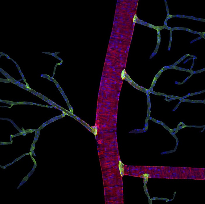
Entry 1: Major artery showing the individual contractile proteins (red) and its nuclei in blue. I chose to enter this image purely for its artistic portrayal of a flower.
Entry 2: The winning image
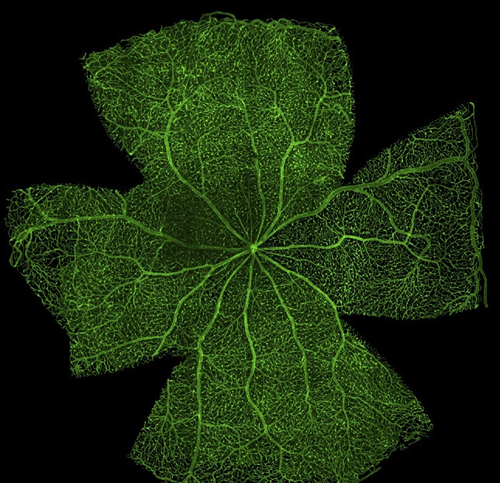
Entry 3: The entire retinal vasculature.
Future Aspirations and Goals:
Looking ahead, what are your key research goals, and how do you plan to build upon your current work in the field of retinal disease?
My key research goals are to identify structural and functional changes in the early diabetic eye that lead to the development of diabetic retinopathy. Structural changes relating to the neurons, glia (supporting cells) and the vasculature are currently being investigated through histological analysis. Functional changes are being investigated my measuring the vasodilatory effect of acetylcholine before and after the chemical induction of diabetes in rodent model. In doing so, I hope to explain what structural changes lead to the functional changes I have already identified in the diabetic eye.
Separate from research, once I have completed my PhD, I hope to attend medical school and eventually work my way towards becoming an Ophthalmologist as it something I have become very passionate about. It is one of the few specialties which incorporates research into direct practise, as I want to continue pursing research.
xxxxx
Conclusion:
In celebrating Hassanain Qambari’s victory in the 49th annual Nikon Small World Photomicrography Competition, we acknowledge more than his visual artistry; we recognize a significant stride in the battle against diabetic retinopathy. His image, capturing the intricate beauty of a rodent optic nerve head, underscores the critical importance of early detection and innovative research in medical science. This accolade not only highlights Qambari’s dedication and the transformative power of combining scientific inquiry with artistic expression but also inspires future exploration and discovery in understanding and overcoming complex health challenges. As we reflect on his achievement, we are reminded of the relentless pursuit of knowledge and the potential of such endeavors to illuminate and change our world.