Astounding Image of Nematostella Vectensis Captured by Ruohan Zhong, A Winner of 2021 Nikon Small World Photomicrography Competition
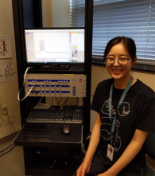
Rooted in childhood curiosity about nature, Ruohan Zhong has devoured knowledge from books, schooling, and research to satisfy her inquisitive mind ever since. Later, Ruohan became an excellent graduate student at Stowers Institute for Medical Research, a cutting-edge research facility focused on foundational biomedical research. At Stowers, she studies Nematostella vectensis, an estuarial creature that has been on earth for billions of years, to unlock the mysteries of their nerve net organization and behavioral implications.
Her recent image of a young Nematostella vectensis was one of the winners of the 2021 Nikon Small World Photomicrography Competition. In the following interview, we have the opportunity to understand a promising young scientist in close-up Q&As. We hope you enjoy reading it.
Q: Congratulations on your recent winning of the 2021 Nikon Small World Photomicrography Competition. Please share with us your path in becoming an outstanding researcher and micrographist.
A: Natural science has been something that fascinates me from the very beginning. I was truly fortunate for having the full support from my parents to binge-watch documentaries about, for example, archaeological excavations or some weird deep-sea creatures near hydrothermal vents, and to have them read “Horrible Science” for me at bedtime before I can read on my own. The attraction never fades with pops of euphoric moments. To name a few: my natural science teacher’s fantastic chalkboard drawing of the subcellular structure of a paramecium first class in middle school; my first time observing pine pollens under the microscope; my first time seeing the “hidden” pigmentation patterns of flowers under a UV camera; my first time hearing about Turing’s theory on the chemical basis of morphogenesis, etc. At the end of college, despite my fondness for life science, I was uncertain whether I could be more than an admirer. Investigator C. Ron Yu at Stowers Institute for Medical Research encouraged me not to rush and offered me an internship opportunity, which eventually led me to the path of graduate school — encounters with so many scientist enthusiasts and their exciting research.
In retrospect, most of the euphoric moments I mentioned above are related to photography/microscopy. In my mind, microscopy is like an alternative window that provides a refreshing view. It allows me to see the strangeness of a familiar object and enhances my appreciation of even the most mundane things. When it comes to my research, microscopy is a helpful tool to solve puzzles, though more often than not, it reveals additional puzzles beyond my wildest imagination. That’s invigorating.
Q: What is your main research interest and why?
A: I am investigating the organization and function of the nerve networks in early-evolved animals. My research interest stems from something that has always intrigued me immensely — the diversity of the nervous system among different forms of life, and their abilities to receive, relay, and process information in a rapid yet controlled manner — as manifested by a coordinated response to the environment. How do complex neural circuits come into being? Does it evolve from a single origin or several independent origins? Does the diversity come from the emergence of novel cellular features or creative ways of using existing building blocks? I believe we can better understand these fundamental questions by a collective of comparative studies of animals from all different evolutionary branches.
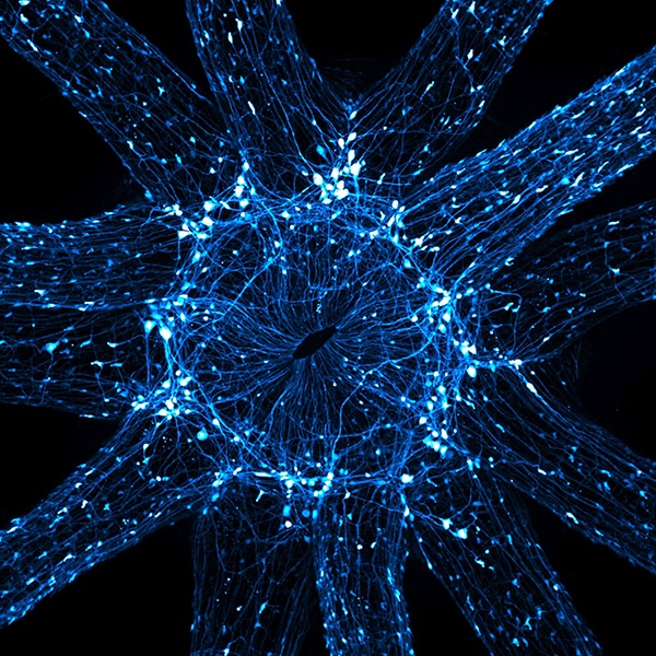
Q: Please share with us the story behind your winning image of “An in vivo snapshot of the neurons surrounding the mouth and tentacles of a juvenile starlet sea anemone”. What type of equipment did you use to take such a brilliant image?
A: Most early evolving animals such as sea anemones are thought to possess a primitive and diffuse nerve net, probably due to their seemingly simple body anatomy and early evolutionary status. However, we still haven’t patched together what their nervous nets actually look like, due to limited tools in neuron visualization. With months of work and luck, I generated a novel transgenic sea anemone with broad neural labeling. For this image, I relaxed a juvenile starlet sea anemone, untangled its 12 tentacles, let it stand upside down, and took a stack of high-resolution snapshots in several seconds before the little anemone lost its balance — all thanks to the ultra-fast imaging by a Nikon Spinning disk!
This image makes me feel that I am standing at a busy interstellar roundabout, surrounded by lightpaths (neurons’ long projections) of passing traffic (the incredible amount of information that is passed by neurons). That was my first impression after taking this image. I was also filled with wonder and excitement because it visually demonstrated various novel neural populations that have never been reported before, which I later learned echoed with some fragmented observations buried in the texts from the Hertwig brothers’ 1879 book (Die Actinien: anatomisch und histologisch, mit besanderer Berücksichtigung des Nervenmuskelsystems).
Q: What have you learned from the above winning image?
A: What we think seems to be inevitably limited by what we can perceive! The nervous net in sea anemones is more complicated than what people have believed previously. Particularly, the clustered neurons at tentacle junctions present an intriguing anatomical structure (ganglia-like). Are they facilitating the coordination between different tentacles? Are they acting as a primitive brain by integrating the information input from all the tentacles, and passing a coherent message to the body columns? This single image piqued myriad possibilities that puzzle and excite me at the same time! Further investigation into the function of those neurons might help us better understand the evolutionary origin of the central nervous system.
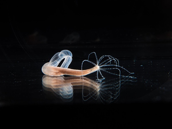
Q: What is Nematostella vectensis? When did they first appear? Why are they important in your research?
A: Nematostella vectensis, also known as the starlet sea anemone, are little translucent creatures who enjoy life between land and sea (brackish water and saltmarshes). They first evolved around 600 Mya, and are among the earliest animals that possess a nervous system. Sea anemones, together with jellyfish and corals, form a clade of animals called cnidarians, which share a common ancestor with bilaterians (all the vertebrates and invertebrates) before the Cambrian explosion. For my research, the sister-group relationship makes them informative slow-evolving models to provide insights on the innovative features that emerged during nervous system evolution.
Among their cnidarians friends, Nematostella acquired increasing attention in the past decade due to their amenability to modern functional genomic methodologies. The transgenesis approach, for example, was how I light up all the neurons with fluorescent protein from the dark unknownness, as shown in the photo. Besides acting as a comparative model to better understand our past, they also exhibit some peculiar features that fascinate me. To name a few, neurogenesis in both germ layers, whole-body regenerative ability, stinging cells that discharge venom through a harpoon-like thread at lightning speed for prey capture…
Q: Besides research, do you mind sharing with us your favorite outdoor/indoor hobbies?
A: I like observing my surroundings, by hiking in nature or simply taking a walk in the neighborhood. It makes me giggle inside when I spot some familiar objectives with a fresh perspective (given the “right” combination of time, lightpath, and my brain). One of my indoor hobbies is classical music and playing the piano. In a sense, that’s an outdoor hobby too because music always takes me to new corners of this world — places that are full of hope.
Q: Is there any scientist or micrographist who inspires your research?
A: I am inspired by Alan Turing and Jean Painlevé. The world in their eyes both represents a sense of Zoological Surrealism (as elegantly put by James Leo Cahill). The former saw simple principles in the chaotic nature of life, while the latter saw chaotic energy beyond our usual still impressions. Both of them inspired me to keep questioning myself — What do I see; what do I think of what I see?
Q: What is your advice for aspiring young people who are interested in fundamental science research?
A: Keep your mind open and gaze around. Let the world surprise you.
*****
We thank Ruohan Zhong for her brilliant explanation of her research topic and interest. We wish her great success in her effort in unlocking the mysteries which are vital in understanding the evolution and innovation of the animal nervous systems.