
Dr. Jianqun Gao’s Winning Image from the Nikon Small World 2022 Photomicrography Competition Helps to Understand Neurodegenerative Diseases
The Nikon Small World 2022 Photomicrography Competition just announced this year’s winners. We are happy to get a hold of Dr. Jianqun Gao to discuss his winning image of Human neurons derived from neural stem cells (NSCs) and his line of research. As a former top neuro medical doctor in Shanghai and a current neurologist/neuroscientist researching at Professor Glenda Halliday’s lab at the Brain and Mind Center, Central Clinical School of the University of Sydney in Australia, Dr. Gao has been devoted to understanding and finding solutions to the difficulties in diagnosis and treatment of neurodegenerative diseases such as Parkinson’s and Alzheimer’s disease.
This winning image was the result of many painstaking trials in culturing Pluripotent stem cells, staining, and image taking. In the end, Dr. Gao was able to capture this clear image by using an advanced Nikon C2 confocal microscope. It helped Dr. Gao and his research team to further understand the mechanism causing Parkinson’s disease and lead to the future development of novel therapeutic strategies for neurodegenerative diseases.
Q: Congratulations on your image of Human neurons derived from neural stem cells (NSCs) winning the top 10 of the prestigious Nikon Small World 2022 Photomicrography Competition. Please tell us about your background and training in becoming an outstanding micrographist.
A: As a neurologist who has been working in a top-level hospital for many years, I have witnessed the development of medicine and at the same time, found various medical issues worth exploring. I want to explore the field of brain science more. Therefore, I completed my Ph.D. research at the Brain and Mind Center, Central Clinical School of the University of Sydney in Australia. The Brain and Mind Centre at the University of Sydney is one of the world’s leading research institutes, where I studied human brain neurons to unravel the underlying mechanisms of neurodegenerative diseases such as Parkinson’s disease and Alzheimer’s disease. It was my great honor to join Professor Halliday’s team. She is a leading top neuropathologist in Australia and worldwide. In the Halliday lab, I learned various techniques utilized in neuroscience research and engaged in exciting research. We were the first to report, using cell models of Parkinson’s disease, that the toll-like receptor 2 (TLR2) exerted its immunological effects on human neurons. We found the target of substance action from complicated signaling pathways and used small molecule inhibitors to inhibit abnormal protein aggregation in Parkinson’s disease. These countless exciting moments motivated me to move forward in scientific research.
This is the fourth time that I won a prize in the Nikon Small World competition for the confocal microscopic images of neural stem cells (NSCs). I would like to thank my supervisor, Professor Glenda Halliday, and colleagues for their continuous support and encouragement. For my research in this area, I was awarded the National Health and Medical Research Council (NHMRC) 2018 Research Excellence Awards, which honors scholars and experts for their outstanding contributions to health and medicine. My confocal microscopic image was selected to be the cover photo of the NHMRC annual report for that year.
Q: Please share with us the story behind this winning image.
A: It is not easy to take this photo. Pluripotent stem cells are a type of cells that are very difficult to culture. Thanks to modern technologies in cell cultures, staining, and imaging, we were able to successfully culture these cells and visualize what was going on in these cells with more detail. Every step of changing the medium, spreading them on coverslips, washing with PBS, fixing with PFA, etc. requires extensive patience and care. In the process of cell culture, in order to make the cells grow better, I have compared a variety of attachment matrices. After many failures, improvement of culture conditions, failures again, and improvement of culture conditions again, I eventually found the optimal conditions for cell growth. In addition, during the whole experiment, I need to explore constantly appropriate experimental conditions, including selection and dilution of primary and secondary antibodies, mounting with fluorescent mounting medium, drying, image capture, and other procedures. Confocal microscopy combined with technologies of digital image processing and electrophysiology enables observation of physiological activities, as well as morphological and dynamic changes, at the cellular level. These advantages make them become important research techniques utilized in morphological, molecular cell biology, neuroscience, and pharmacological research.
Q: What are neural stem cells (NSCs)? What is the main goal of your research by studying NSCs?
A: Neural stem cells are stem cells within the nervous system that can self-renew and evolve into differentiated precursor cells that give rise to various types of cells such as neurons and glial cells. I am studying the formation of pathological α-synuclein inclusions in human neuronal cells induced by exogenous α-synuclein fibrils, resulting in impaired autophagy and increased release of abnormal α-synuclein species that may affect other neurons. Meanwhile, I am also studying the contribution of neuronal toll-like receptor 2 (TLR2) to the spread of pathogenic α synuclein. My research interest stems from a topic that has always been of great interest to me: why are people getting old? I believe that research on these disease models will allow us to better understand these fundamental questions. Our primary goal is to understand the mechanisms by which the brain undergoes self-clearance and repair; and also to help design specific therapies aimed at improving neurological function and quality of life in patients with CNS injuries and neurodegenerative diseases. By using these disease models of NSCs, we will be able to investigate the potential of certain small molecule compounds as PD drug candidates and help patients in the future.
Q: What have you learned from this image and how does it help you understand the molecular and cellular properties of human brains?
A: Neural networks and nerve cells are much more complex than we think. This picture sparked my reverie of countless possibilities, leaving me both bewildered and excited. Further studies of the function of these neuronal cells will help us better understand the occurrence and progression of CNS diseases. As a neurologist and neuroscientist, I am focusing more on the neural pathways of the brain and central nervous system.
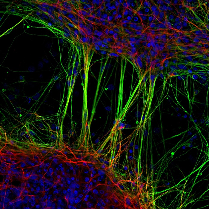
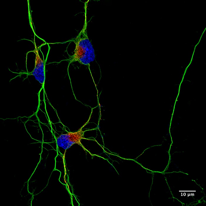
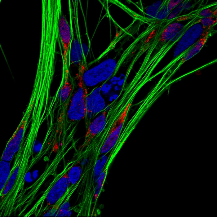
In this image, these human neurons are differentiated from neural stem cells (NSCs). Cells have been stained with markers that depict the nuclei of neurons (blue) and their networks (green), abnormal protein aggregates (yellow), and autophagy mechanisms (red). I used immunocytochemistry in my research: first treating these neurons with activators to induce the formation of abnormal protein aggregates (which is to generate a cell model of Parkinson’s disease), and then treating the cells with inhibitors (which is to observe whether it may be a potential drug candidate). By conducting research on these NSCs, we hope to unravel the mechanisms and develop novel therapeutic strategies for neurodegenerative diseases, such as Parkinson’s disease.
Q: What equipment and technology did you use to take such crisp, clear images of NSCs?
A: I captured this clear confocal microscopic image with a Nikon C2 confocal microscope. When taking this picture, I used a red fluorescent dye to label neural stem cells (NSCs), a green fluorescent dye to label abnormal α-synuclein accumulation, and took a bunch of high-resolution photos, all thanks to the power of confocal microscopy! Imaging technology is not just a technique, but a powerful tool for deep exploration of the unknowns.
The preparation of samples, the selection of color developers, the rational combination of laser and color developer irradiation attenuation, and the balance of spatial and temporal resolution all contribute to the achievement of a high-qualified picture combining science and art. The quality of traditional fluorescence microscopic image is not high, which makes tiny changes difficult to observe. Confocal microscopy has provided the solution to the problem of blurred focus, which allows us to recognize these tiny structures in a clearer way.
This photo reminds me of the sandwich I eat, with two slices of “bread” on top and bottom, and the filaments in the middle are like heated “cheese”, which is my first feeling after taking this image. In addition, this image also surprised and excited me simultaneously, because it visually shows the changes in Parkinson’s cells after drug treatment, and a confocal microscopy is a powerful tool for the exploration of the micro-world.
Q: By identifying biochemical signatures in the brains of patients with neurodegenerative diseases, what kind of applications or treatment plans can be derived from that?
A: By showing the dynamic relationship between the accumulation and degradation of α synuclein inclusions in human neuronal cells, and the involvement of TLR2 signaling in α synuclein accumulation and pro-inflammatory response in human neurons, we aimed to provide evidence for targetable mechanisms to intervene in the pathological progression of Parkinson’s disease (PD). PD is a progressive neurodegenerative disorder that affects approximately 7 million people worldwide. Its pathological characteristics are the loss of dopaminergic neurons in the substantia nigra pars compacta region of the brain, accompanied by the formation of Lewy bodies, and the protein α-synuclein is the most prominent component in the pathogenesis of PD. My work has also provided proof of the principle that targeting these mechanisms shows some promise at alleviating at least a proportion (or more) of the pathology. Therefore, the research model established in my work could potentially be utilized further for PD research. The data support the use of autophagy enhancers and TLR2 antagonists for ameliorating α-synuclein accumulation, but further work is still required. My work provides a strong foundation for future studies investigating the consequences of α-synuclein and TLR2-mediated pathology in PD, and also for finding novel strategies for the treatment of PD using these small molecule compounds.
Q: Besides researching at the lab, what other hobbies or activities are you fond of?
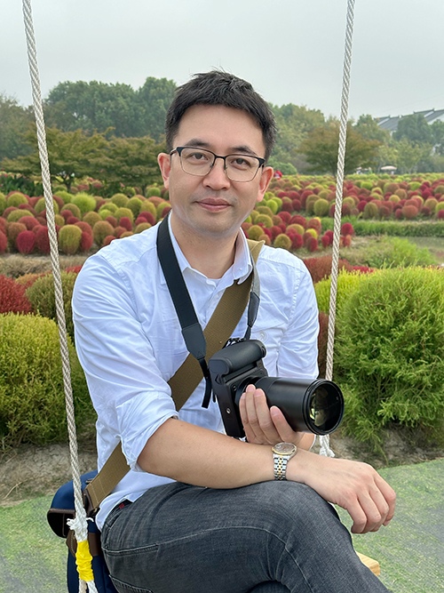
A: I am an amateur photographer who likes to travel around the world to enjoy beautiful scenery and record my feelings with my cameras. I am particularly sensitive to colors, which means I can tell subtle color differences with my naked eyes, which is probably one of the reasons why I am obsessed with photography. My photos have been selected to be used on the official website of the New Zealand Tourism Organization.
Q: Is there anything else you would like to tell our readers?
A: The exploration of disease mechanisms and treatment for neurodegenerative diseases is still a long journey. We feel grateful to the patients, healthcare workers, researchers, social workers, and others for their enthusiasm and willingness to participate in this journey. Advanced technology has facilitated and helped us along this path, enabling visualization of what was difficult to achieve in the past, especially the micro-world, and helping us to learn more about the field. If you are also interested in neuroscience, please persevere and keep exploring, because the little knowledge we gain will bring vitality to the difficult and profound ocean of brain science.
*****
Dr. Gao’s winning image is a testament to the importance of microscopic technology which helps many people in understanding nature. We appreciate Dr. Gao spending the time to share the story of his winning image and the research he is involved with. We look forward to more exciting news from him in solving the mysteries of human neurodegeneration.
Dr. Gao’s contact information:
LinkedIn: Jianqun Gao
Facebook: Jianqun Gao
Twitter: @drgaojqun