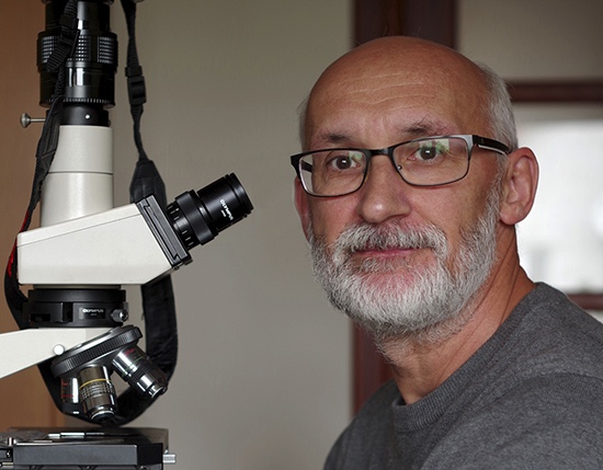
Introducing the Extraordinary World of Photomicrography: An Interview with a Microphotographer, Marek Miś
Marek Miś, a biologist with an unwavering passion for photomicrography, discovered his calling, capturing the hidden wonders of the microworld. Inspired by the book “Microbe Hunters,” Marek embarked on a journey to explore and document these hidden realms through his camera lens. Photomicrography, a challenging yet rewarding form of photography, demands technical expertise and an understanding of microscope operation. Marek overcomes obstacles such as specimen preparation, sample selection, and illumination techniques to unveil the astonishing beauty and diversity within the microcosm, granting others the opportunity to marvel at these hidden worlds.
Marek’s exceptional work in photomicrography has earned him international acclaim, with his photographs featured in renowned competitions, exhibitions, and publications worldwide. From esteemed contests like Nikon Small World to prestigious exhibitions such as “See Me – The Story of The Creative” and “Beyond the Bulb,” Marek’s captivating micrographs have gained widespread recognition. Through his distinct artistic style, Marek aims to convey his passion and unveil the mesmerizing micro universe to a broader audience. In the following interview, Marek shared with us the journey of becoming an outstanding Microphotographer. We hope you enjoy reading it.
Q: Please tell us about your background and how you became interested in photography, particularly in the field of photomicrography?A: I am a biologist by education. I graduated in biology at the Nicolaus Copernicus University in Toruń. Nature in its various forms fascinated me from an early age. I don’t know why, but I’ve always been especially interested in the world of small things; I was interested in insects, I looked at the details of the structure of flowers. I received my first camera as a gift from my father when I was 12 years old. Of course, I mainly photographed nature, in particular insects and flowers. This is how I started my adventure in photography. At that time, I didn’t even think about microphotography, I didn’t even have a microscope.
Q: What inspired you to specialize in photomicrography and explore the world of tiny things?
A: My interest in the world hidden from the unaided eye began quite by chance. When I was 13 or 14, I helped my father clean out his closet. That’s when I came across an old, tattered book by Paul de Kruif called “Microbe Hunters”. What I read changed my life. It was then that the passion for discovering microworlds was born in me. I was especially fascinated by Antoni van Leeuvenhoek. I was impressed by what he accomplished with simple hand-made microscopes. I wanted to discover the mysterious world of microbes, plants, microscopic animals and everything that fell into my hands in a similar way. The lack of a microscope was an obstacle. At that time, in my country, it was practically impossible to buy a microscope; they were not available in stores. And even if they did, my parents weren’t able to afford one anyway. Therefore, like Leeuvenhoek, I decided to build my first microscopes myself. I was grinding my own lenses. I also used the bottoms of tiny bottles used for cake scents as lenses. Later, I used the lenses of small magnifying glasses to assemble my first compound microscopes. I used wood, metal, and cardboard to build them. The magnifications were not great, but they allowed me to explore the microcosm for the first time. I remember very clearly how impressed I was with the first observations of snail embryos rotating inside egg shells, the first observations of protozoa, or the anatomical structure of various plants. Since I really liked to draw, I immortalized everything I saw on paper using pencil drawings. I wanted to record what I saw for myself, but also to show this fascinating world to others; photos instead of drawing. This is how my adventure with microphotography began. I wanted to record what I saw for myself, but also to show this fascinating world to others. I still have these drawings today 🙂
When I was a bit older (I must have been 18 at the time) I thought about taking photos instead of drawing. To achieve this, I destroyed my first camera (equivalent to today’s compact cameras), depriving it of the lens. My first microphotographs, which I took in 1980, were black-and-white and of frog blood. I have that negative to this day. This is how my adventure with microphotography began. As I was not using a SLR at the time, it was very difficult to obtain technically correct microphotographs. Everything was based on trial-and-error. This required the use of more photosensitive materials than usual. Because at that time in my country there were great difficulties with the purchase of photographic materials, and when they were available, they were prohibitively expensive, I gave up this type of photography for many years. I returned to it only when I bought my first digital camera in 2009. It was a Pentax K10 camera. Since 2009, microphotography has absorbed me completely. The enormous possibilities of digital photography meant that nothing stood in the way of devoting yourself to it completely.
Q: How would you describe the unique characteristics and challenges of photomicrography as a form of photography?
A: Microphotography is a special and difficult field of photography, because apart from generally good camera skills, it requires excellent knowledge of the operating of the microscope and the use of its capabilities. Good microphotography also requires knowledge of how to use the available methods of illumination of the specimen to obtain a satisfactory result. Many factors determine the final result. It is very important to properly prepare the appropriate preparation. This is the starting point for good microphotography. In addition, it is very important to know how to obtain materials for a specific category of photos. You have to be able to take the right samples. It is also important to store them properly to be able to photograph them for a long time. The microscopic image is characterized by a very small depth of field, which is a big problem when we want to photograph an object, so that all the details are sharp in the photo. That is why the stacking method is necessary. Another problem is the vibration taking place while taking the picture; it is a big challenge avoiding them. Even the smallest vibrations, when dealing with significant magnifications, can result in blurred photos.
Q: In your opinion, how does photomicrography allow you to discover and reveal hidden worlds to others?
A: Looking through the eyepieces of a microscope at a sample for the first time is the thrill of discovery. Of course, I usually don’t discover anything new at all, but I certainly discover many things for myself for the first time. Usually, I want to share my “discovery” with others. I do this through my Facebook and Instagram accounts, as well as on my website. People are generally unaware of the beauty and great diversity of the microcosm. Thanks to my photographs, I want to show them at least a small fragment of this fascinating reality.
Q: Could you share some insights into the techniques of illumination you use in your photomicrography and how they contribute to the richness of colors in your micrographs?
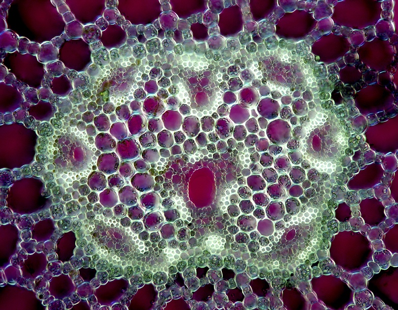
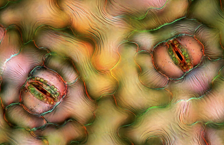
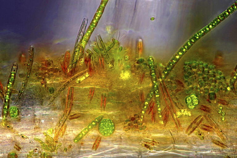
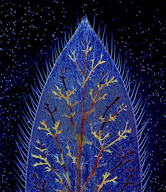
A: I use several techniques of illumination: bright field, dark field, polarization, Rheinberg, phase contrast, and DIC. My favorite techniques are polarization and dark field; I combine them both very often. After taking photographs I usually improve them by slightly increasing the contrast and sharpness. Apart from that I check every photo at the magnification of 100% and remove dust from the sensor. That is all. I never change the colors. For polarization and dark field I use homemade accessories.
Q: What kind of equipment do you use for your photomicrography, including your choice of microscope and cameras?
A: When it comes to the microscope I use an Olympus BH-2 BHS equipped with a 100W halogen lamp. For the objectives I use Olympus Splan Apochromats. I photograph with a Pentax K-1 DSLR and lately with a Sony Alpha 7 R IV mirrorless camera. Both cameras are the full-frame ones. The cameras are connected to the microscope with a special photo tube. The microscope and photo tube were bought on eBay. I’ve been completing my microscope for several years. For the phase contrast I use the original Olympus device. But for DIC (differential interference contrast) I use DIC parts from a Polish microscope, which I had to modify a bit to fit it to my Olympus.
Q: Your micrographs have been published in various publications and exhibited internationally. Please share with us some highlights of your achievements and experiences in showcasing your work?
A: Indeed, there are several international exhibitions where my photographs have been shown. I am particularly proud of the group exhibition in New York called “See Me – The Story of The Creative” in 2013. It was also very important for me to show three of my works at the worldwide traveling exhibition organized by UNESCO in 2015 called “Beyond the bulb”. The exhibition was shown in many countries, on all continents as part of “The Year of Light” celebrations. Many of my micrographs were also shown internationally after various competitions as the part of the exhibits (e.g. Nikon Small World, Olympus BioScapes, and others). The international exhibitions are the opportunity for showing my way of seeing the microworld. As for publishing my works in various publications, they are used all over the world thanks to my collaboration with specialized photo agencies: Science Photo Library (UK) and Science Source (USA). Apart from that, I offer my works as high quality prints to individual clients on Etsy and on my website. This is an additional way for showing my works internationally 🙂
Q: How do you approach photomicrography as both a scientific document and an art form? How do you convey your feelings and capture the beauty of the micro universe in your images?
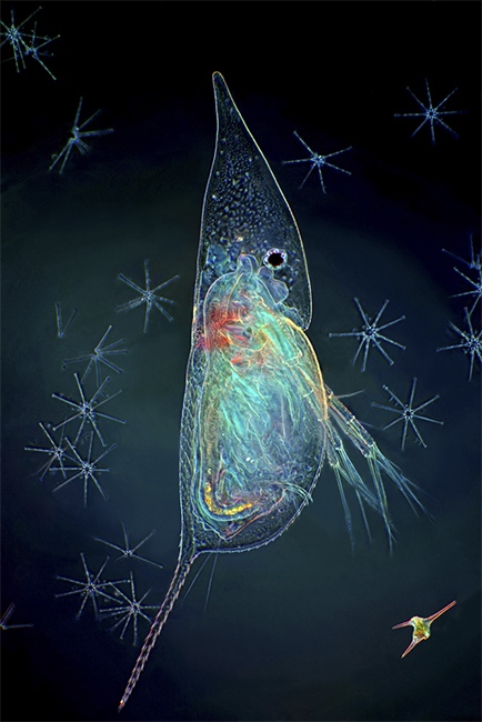
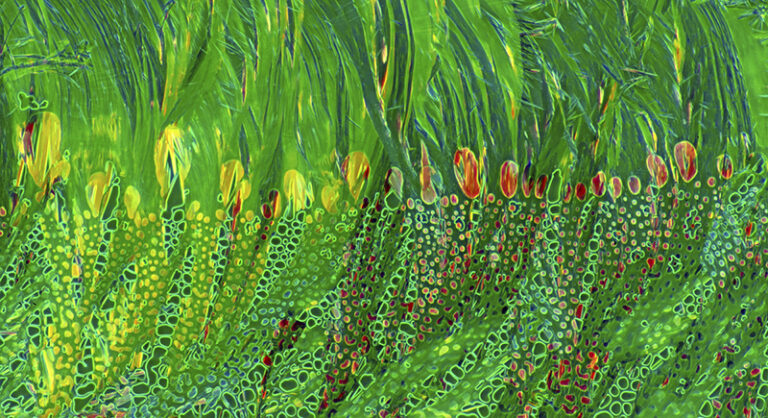
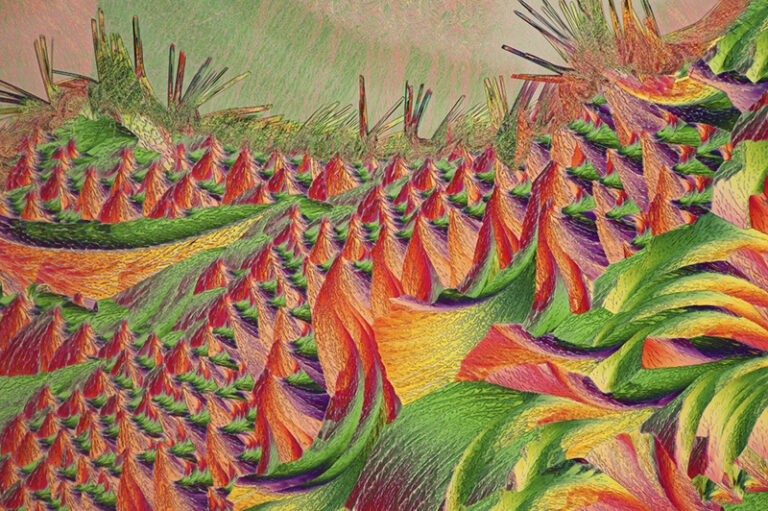
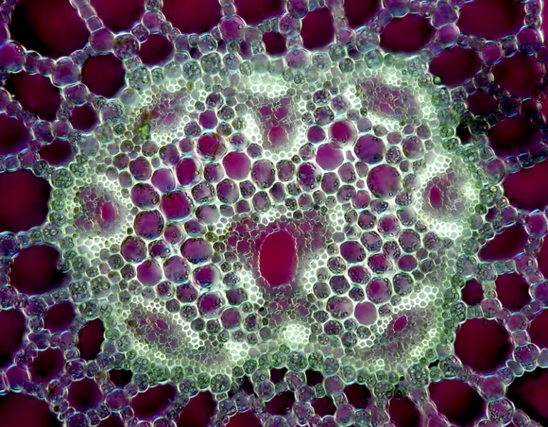
A: Although I am a naturalist, I also really like to photograph objects completely unrelated to nature, such as air bubbles or various recrystallized substances. Photographing these substances gives me a lot of joy, because it allows great creative freedom. I always try to make my photographs more than just a raw record of what I see under the microscope. Even if it is a purely biological object, I always try to photograph it in such a way that it looks interesting. To achieve this, I check how it will look in different lighting, in different positions, and finally in a different environment. I usually try to photograph a natural object not in isolation, but in the company of other organisms and structures from the same sample. In this way, I photograph it, as it were, with a part of its natural environment. Then something happens in the photo, it becomes more alive and dynamic. Of course, I also photograph isolated objects because I know that there is often a demand for this type of photography, especially in various scientific publishing houses. However, photographing the entire microscopic particle of the microworld, not individual organisms, gives me the most satisfaction. I probably have an artistic soul, that’s what others say about me, so I guess that’s the reason for my artistic approach to microphotography, ha, ha, ha 🙂 It’s always a huge compliment for me when someone says that by looking at my photos they immediately know that I am their author, that I have my own unique style 🙂 This is very important for a photographer with an artistic soul. It is very important for every artist to refine their own style. But it’s not planned. This happens by the way, naturally. This cannot be programmed and is probably the essence of the artistic approach to photography. You just have to have it in you and it shows up in what you do, be it painting, sculpting, photography, or any other field of art.
Q: You have been recognized in prestigious photomicrography competitions such as Nikon Small World and Olympus BioScapes. How does it feel to be acknowledged for your achievements in this field?
A: Contests are something that allows one to compete against others in a certain field. Thanks to them you can see what others are doing. You also see how they do it, what topics they choose, how they approach them, and what methods they use. This is very developing. Seeing so many great photos in one place is very inspiring. Looking at a very good photo taken by someone else, sometimes you wonder why I didn’t come up with a similar idea, why I didn’t choose this or that subject. Others, who are looking at your photos, probably think similarly 🙂 That’s why competitions develop all participants and it’s worth taking part in them. Of course, everyone who participates in the competition wants their work to get the best result; it’s normal. Winning, or at least qualifying for the top, gives great satisfaction and motivation to further improve one’s skills. I agree with the saying that my best picture is the one I haven’t taken yet. This approach forces you to constantly work, to constantly improve in what you do. The thing about contests is that sometimes you win and sometimes you don’t. Therefore, it is much more important to me that the organizers of the Nikon Small World competition, in recognition of my achievements so far, have included me in the respectable group of Masters of Microscopy. I am additionally satisfied with the fact that I am in this group as the third in a row 🙂 I am equally satisfied with the fact that last year, as one of the few so far, I was awarded by the Science Photo Gallery agency with a special interview and the term Master of Microscopy. Awards of this kind are probably more important to me than winning competitions.
Q: Beyond your photography, you also conduct lectures, workshops, and exhibitions related to the micro world. Please tell us more about these activities and your efforts to educate and inspire others.
A: Currently, my main activity popularizing knowledge about the microworld and the ways of capturing its beauty with photography are special workshops that I run at my home. These are individual workshops in which only one person participates. This allows for greater efficiency of such workshops. Earlier, for several years, I also conducted group workshops in a cultural center in my city. My workshops are attended by people of all ages ranging from teenagers to adults. I always get great satisfaction when I see their enchantment with what they can see under the microscope. They are always simply delighted and amazed at the great diversity of this tiny world. I can also boast that I had participants not only from my country, but also from the Netherlands and the USA 🙂
In addition to workshops, I also conduct, from time to time, shows of my photomicrographs with commentary in schools, both primary and secondary. In this way, I try to popularize knowledge about the microworld. The rich colors of my photographs help me a lot in this. It shows this world, which is quite little known to young people, in a completely different, attractive way than in textbooks. My solo exhibitions play a similar role. By showing the microworld in an attractive way, I believe that I contribute to arousing more interest in it in the viewers. Usually during exhibitions I am asked about how I took this or that picture. Some people seem to be so fascinated by it that they ask about taking part in my workshops 🙂 I also popularize knowledge about the microworld and microphotography through various articles in magazines, as well as by giving interviews such as this one. In 2015, I also wrote a book half devoted to this subject: “Blisko, coraz bliżej. Od fotografii zbliżeniowej do mikrofotografii” (Helion 2015). I dream of writing a book devoted exclusively to the microcosm and microphotography. I would like it to be available also in English, so that I can share my experience not only with my compatriots.
In conclusion, I am immensely grateful to Marek Miś for participating in this interview and sharing his remarkable journey as a micro photographer. Marek’s passion for capturing the beauty of the microworld through his lens, coupled with his technical expertise and artistic vision, allow us to witness the awe-inspiring wonders that exist beyond our naked eye. His dedication to photomicrography serves as an inspiration to aspiring photographers and scientists, encouraging them to explore the hidden realms of the microcosm and share its extraordinary beauty with the world.