Hebe plant anther with pollen (magnification 100x), Laser confocal microscopy by Robert Marku, Biologist, Micrographist, UK

Stunning Images of Nature by Dr. Robert Markus

Winner of 2020 Nikon Small World Photomicrography Competition

We are honored to be able to interview Dr. Robert Markus, winner of the 2020 Nikon Small World Photomicrography Competition. His winning image, “Hebe plant anther with pollen”, displays crisp details showing how cells of the Hebe plant react to light. His procedure provides vital information needed for understanding the plant at its microscopic level. 

Robert, as an experienced micrographist, has been looking at different objects from nature in fine details and has produced breathtaking images. Robert uses a sophisticated laser confocal microscope and plays with lights with a spectral detector instrument to get amazing results.  These images not only have yielded significant scientific findings, they are also highly regarded for their aesthetic presentation. 

Robert’s candid responses in the following interview will shed light on the process he uses to produce his winning image and his personal interests in some of the projects that he has been conducting currently. Owing to Robert’s expertise in utilizing advanced scientific instrumentation, we are better able to see what an amazing world we live in.

Q: Your “Hebe plant anther with pollen” image won the 2020 Nikon Small World Photomicrography Competition. Congratulations! Please tell us more about this image and the technology you employed in producing such an amazing image.
A: Thank you for your kind words.

The image shows a microscopic view of an anther with pollen, the part of the plant that holds the genetic material locked inside the pollen, the small round, colorful ball-like structures in the image. Plants display a high variety of forms and shapes for these reproductive parts and show variable reflectance and absorbance of light. This feature can be explored using UV light, and so calls for a so-called laser confocal microscope. We illuminate the sample with blue (UV) and we record the reflected and emitted light. Because plants contain many “colorful” molecules that can absorb light. These molecules can change the nature of the light / the color is detected as a mixture of colors which can be separated with specialized instruments (so-called spectral detector) attached to the microscope; we use this for research usually. Taken with artistic freedom, we combine the various spectra and recolor the data. Additionally, we aim to represent the naturally occurring colors which we would see if we would use a UV torch to illuminate a plant and look at it with various filters. The scientific instrument (laser confocal microscope) makes this process more accurate and at a much higher resolution than we could achieve with regular macro photography. The image does not directly relate to the research we do, however, the sample it is made from is a plant (Hebe bush) growing on the campus. To make awareness of the natural world and beauty around us we search for samples to scan using the above-explained technique.

Hebe plant anther with pollen (magnification 100x), Laser confocal microscopy by Robert Marku, Biologist, Micrographist, UK
Hebe plant anther with pollen (magnification 100x), Laser confocal microscopy. Courtesy: Robert Markus and Zsuzsa Markus

Q: Please share with us the path you followed that led to you becoming a top micrographist.
A:
As a child, I was intrigued by Jacques Cousteau’s documentaries on ocean life, which was to me the perception of research and exploration; and I liked optical devices, telescopes, and microscopes, so microscopy was the way to explore the world around me. My parents always helped me to explore science and helped me to build things, bought me very useful books (well before the internet existed, and I grew up without computers and smartphones). In those times, we had books, microscopes, sports, and hiking to entertain ourselves, and my father liked photography too, and I liked landscape and macro photography. I always liked biology and science and dreamed of being a scientist someday. Teachers, mentors helped to choose the path to be a biologist. Later on, research became feasible and I was fortunate to do a Ph.D. in biology and became a postdoctoral fellow in various countries. Technically, I wanted to know how microscopes and cameras work, and that passion helped me to learn and, in the end, usually I was assigned to be the person taking care of microscopes at the departments I worked. In the last six years, this became my main role, I am a Senior Imaging Technician, and super-resolution microscopy scientist. Thus, in the last 20 years, I am working daily with microscopes and in my free time, I do explore samples and search for beautiful things to photograph.

Q: What objects interest you the most when producing micrographic images? What are you looking for; the aesthetic aspects or scientific findings?
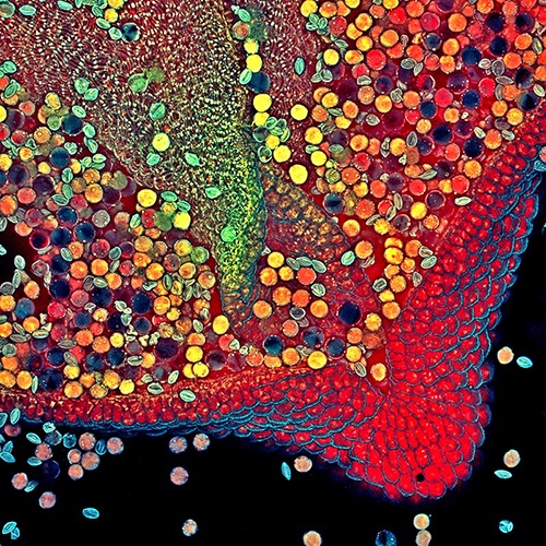
A:
My scientific curiosity is fulfilled daily at work by working on various projects. For artistically pleasant and visually impactful images I do search for subjects. Plants, flowers especially are amazing and there is still a lot out there to explore. I explore the visual impact of a subject, I work on the color composition and framing, and I do repeat the “visual experiments”, I go back several times to the same subject to aim to make better and better images.

Q: Lighting techniques are vital in producing crisp, clear micrographic images. Please share with us some of your experiences when using lighting in atypical ways to yield different interpretations.
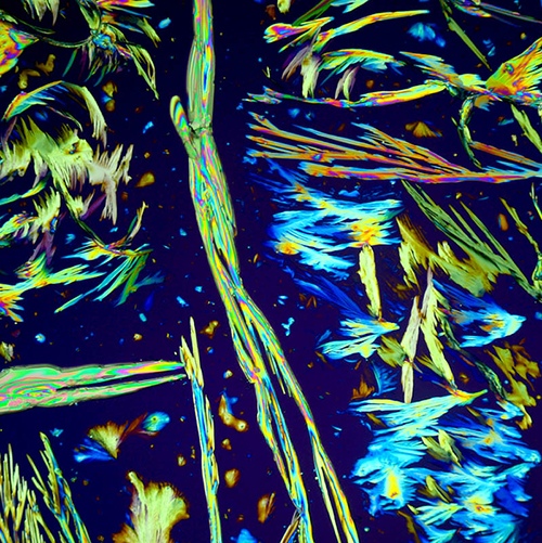
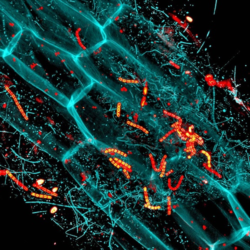
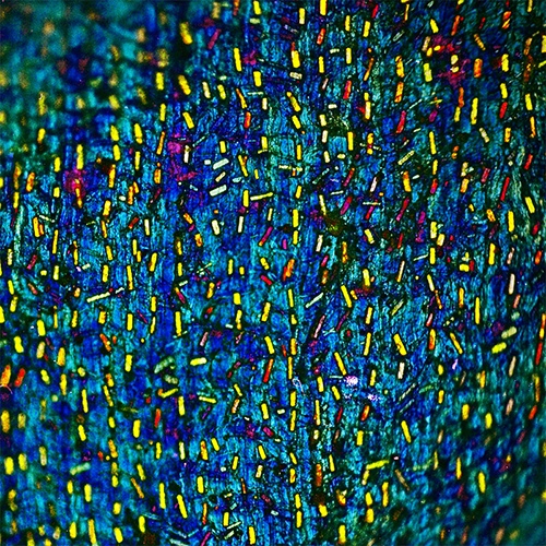
A:
Usually I work with the laser confocal microscope, which uses a small aperture to produce digital images with good contrast and sharpness. By scanning slowly we allow the system to reduce the detector noise, that is one of our extra tricks to achieve the best we can get out of the instrument. For other types of illumination, I use side illumination or back illuminate samples (color dark field), or many times I use at home a modified vintage microscope to create colorful images of crystals, using polarization filters. Additionally, drift or camera shake needs to be avoided, with DSLR I use mirror lock-up, or I use a small mirrorless camera and electronic shutter only. Back in the old days, I adapted flash photography on my home-built microscope system.

Glycine crystals under polarized light, using a modified vintage microscope and DSLR. (magnification 100x) by Robert Marku, Biologist, Micrographist, UK
Glycine crystals under polarized light, using a modified vintage microscope and DSLR. (magnification 100x). Courtesy: Robert Markus

Q: Please share with us other examples of your image production that led you to unexpected discoveries.
A:
The other image submitted to Nikon Small World 2020 explores the decay of a duckweed root. That was originally a sample used to view single-cell organisms in the pond. I left the sample on the bench and looked at it again after a week and observed it was hazy. The reason for that became obvious when I explored it at various magnifications, and took many images of bacteria growing in the sample and which started to transform and degrade the organic matter.

Decay of duckweed (magnification 400x) by Robert Marku, Biologist, Micrographist, UK
Decay of duckweed (magnification 400x), microorganisms are red and cyan, and the cell wall of the duckweed (large brick-like structures) are cyan too. Laser confocal microscopy. Courtesy: Robert Marku

Q: If you don’t mind, please tell us about some projects that you are currently working on.
A:
For nostalgic reasons, I started to explore film photography again, and I made a few images of onion skin under polarized light. Next was to create a nice image of the garlic skin. I am trying to make a series of images that would go well together, exploring the vegetables in the kitchen. However, flowers look exotic under the microscope, and always hold visual surprises, so I am waiting for the spring to explore the newcomers.

Onion skin under polarized light, analog film photography using a modified vintage microscope, and an SLR from the 1980s by Robert Marku, Biologist, Micrographist, UK
Onion skin under polarized light, analog film photography using a modified vintage microscope, and an SLR from the 1980s. The colorful regular stone-like shapes are oxalate crystals, which serve as a defense against animals eating the bulbs because it causes slow digestion. (Magnification 100x). Courtesy: Robert Markus

Q: In your opinion, what does it take to be a good micrographist?
A:
Passion, perseverance, very good knowledge on how to use the microscope you have, even better if one fully understands how it works, know where and how to work with the light, sample preparation is crucial. Additionally, one needs to study arts, visual expression, don’t leave it to luck, do explore more, and discuss with artists your creation. I am very fortunate to have my wife with a biology background and her passion for painting and drawing. We make a good team to create the images we like.

Q: Over the years, you have captured many wonderful images of various species in nature. From these observations, what is Nature to you and what have you have learned from it?
A:
I consider Nature as a gift; we are privileged to witness it, and we should not harm it. I know, there are demands, and humanity needs to modify certain landscapes but using science we should do it in a sustainable way. When I think of the rocks, the stars, and the universe we know, nothing nearby is as special as Planet Earth. I hope we as humanity will be able to protect it, and not waste it. Microscopy opens another world with shapes, structures, and complex relations between molecules, cells and eventually, our visible world is bound to those forces at a microscopic level too. Exploring the universe tells us how precious Nature and the life on earth is.

Macro photo of moss after the rain by Robert Marku, Biologist, Micrographist, UK
Macro photo of moss after the rain. Courtesy: Robert Markus

We thank Dr. Robert Markus for sharing his images as well as the stories behind these images. His close up images of nature help us to see these tiny but important creatures that help to sustain this planet earth. Want to find out more stunning images from Robert, you can check out the link below.

Robert’s Website