Winner of 2021 Nikon Small World Photomicrography Competition

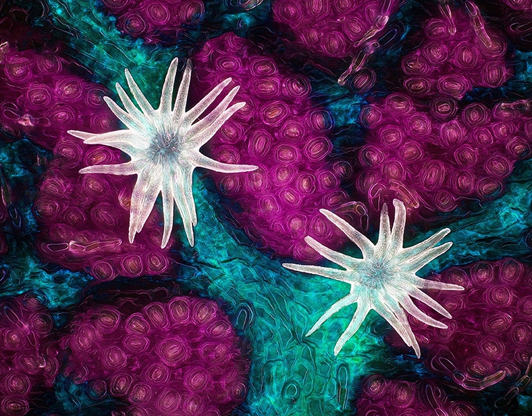
It is our great honor to be able to interview the winner of the 2021 Nikon Small World Photomicrography Competition, Jason Kirk. Jason is the Director of the Optical Imaging and Vital Microscopy Core facility at the Baylor College of Medicine. His “Trichome (white appendages) and stomata (purple pores) on a southern live oak leaf” image won first place while his other image, “Vasculature of a mouse retina,” won 11th place in the same competition.
For his “Trichome (white appendages) and stomata (purple pores) on a southern live oak leaf” image, he used 60X magnification and stacked more than 200 different images to see the structure in depth. The image displays three different leaf structures: stomata (purple), trichomes (white), and veins (blue). This image demonstrates that something as simple and as common as an oak leaf consists of uncommon beauty and intricacy.
Jason has been consistently creating astounding images by exploring the very tiny and vital structures in nature. These images not only provide amazing visual effects but also evoke people’s curiosity about the roles these structures play in facilitating life, not to mention encouraging further research on them.
In the following conversion, Jason shares with us his path to becoming an outstanding micrographist and his stories of some of his winning images. We hope it inspires you to learn more regarding the beauty and vital function of the structures that can be seen at the microscopic level.
Q: Congratulations to you on your winning of the 2021 Nikon Small World Photomicrography Competition. Please tell us about the path you took to be such an excellent microscopist.
A: My interest in microscopy started by accident. While an undergraduate at the University of Maine in the mid-’90s, I fell in love with photography after spending a summer studying herpetology in Costa Rica. When I returned home, I spent much of my time learning how to be a photographer and eventually joined the staff at our campus newspaper. To get pictures published, they had to be turned around quickly. This required a darkroom, for which the newspaper had only one. My advisor in the biology department, Dr. Seth Tyler, had a darkroom as part of his electron microscopy facility, and in exchange for work-study, he agreed to let me use the darkroom whenever it was available.
It was in this lab that I saw my first microscope with a digital camera attached to a computer. This technology was mind-blowing at the time, and I had to know more. Dr. Tyler suggested I take his graduate-level electron microscopy class where I learned basic light microscopy as well as electron microscopy.
Once I completed my degree program in 1999, I used this experience to get a job at the Marine Biological Laboratories in Woods Hole, MA. I spent half a year working for Carl Zeiss supporting a huge variety of light microscopes for the summer courses held annually at the MBL. This is where I first started to use fluorescence and confocal technologies that I still use to this day.
My experience at the MBL kickstarted a career in microscopy. From MBL I spent four years at the University of Connecticut Health Science Center, running the day-to-day operation of the CBIT Microscopy Facility. Under Dr. Leslie Loew and Dr. Ann Cowan, I learned a ton about cell biology, fluorescence techniques, and confocal/multiphoton microscopy. After UCHC, I spent 12 years working in the advanced imaging group at Carl Zeiss Microscopy before moving to the Baylor College of Medicine in 2017 to direct the Optical Imaging & Vital Microscopy core facility.
Q: Your “Trichome (white appendages) and stomata (purple pores) on a southern live oak leaf” image won first place at the 2021 Nikon Small World Photomicrography Competition. Please share with us the story behind this image and the techniques you used when taking this crisp, beautiful image.
A: Before COVID hit the world, I had been building a small microscope designed to join my SLR cameras with microscope objectives. When we were quarantined for a few months in early 2020, this project accelerated, and I started looking for subjects to test it out. My daughter brought a bunch of different things from our yard to observe, and one of them was a leaf from this tree.
The southern live oak (Quercus virginiana) is a common tree found here in Texas, and I was amazed by the amount of trichomes growing on the underside of this leaf. My daughter remarked that they looked like sea anemone, which sparked the idea for this image. After a bit of research on trichome function, I waited until the following spring when the leaves were just sprouting on this tree. During these early stages, trichomes are not as dense and this allowed me to locate a few newly formed structures for this composition.
The image was created using diffused reflected light to illuminate the trichome as well as transmitted light filtered through a red filter to colorize the veins. The original colors in this image were green surrounding the white trichome with red veins. The colors were shifted in Photoshop to create this undersea color palette.
Q: As a director, please tell us about the main tasks of the Optical Imaging and Vital Microscopy Core facility at the Baylor College of Medicine and the applications that have been developed there?
A: As a core director, it is my job to facilitate research using microscopy. Microscopy is an essential tool for many scientific research disciplines, and these instruments tend to be expensive and complex to use and maintain. In addition to caring for the equipment, we instruct a wide range of investigators on how to properly utilize these instruments to gain useful insights into the biology they study.
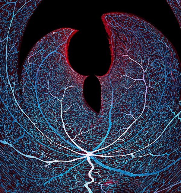
Q: Please tell us about your other winning image “Vasculature of a mouse retina”. What have you learned from this image?
A: The morphology of blood vessels is an important aspect of understanding how vertebrate embryos develop. These mouse model systems drive basic science research toward a deeper understanding of how these vessels grow, repair themselves and respond to disease or genetic anomalies. This research has ripple effects and feeds every avenue of clinical treatment for human disease.
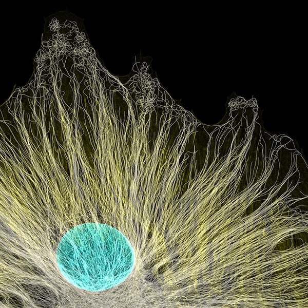
Q: Your Image of “Microtubules radiating from a tissue culture cell” won the Image of Distinction in the 2020 Nikon Small World Photomicrography Competition. It beautifully displays the microtubules within a cell. Please tell us more about this image and why you were attracted to microtubules?
A: You said it – microtubules are simply beautiful. They form these mesmerizing patterns within cells and constantly keep me in awe. You are hard-pressed to find a better subject.
Q: Please share with us some of the projects that you are currently working on.
A: I am working on perfecting a microscope to perform high NA imaging on large image sensors. This requires optics and components that can handle projecting an image uniformly over a large field number >30mm.
Q: For aspiring micrographers, do you have any advice you would like to share?
A: Find mentors. Identify people you admire in this field and don’t be afraid to contact them with thoughtful questions. I know so many amazing microscopists that are generous with their time and knowledge that would happily invite conversation about microscopes!
*****
We appreciate Jason’s time and effort in replying to our questions. His candid words and images definitely instill our appreciation and desire to learn more about nature.
Jason’s Website, Instagram, Twitter: @jasonmileskirk