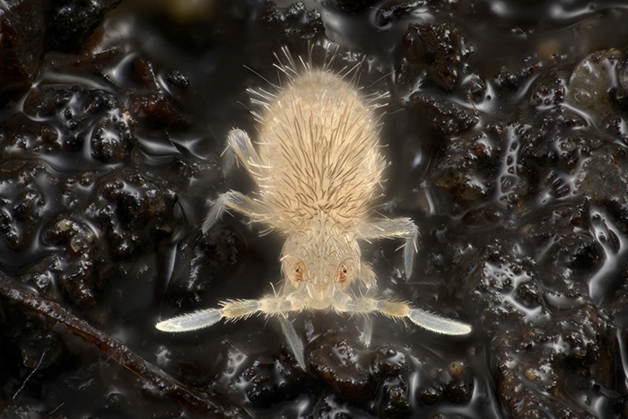
The Microscopic Muse: Wim van Egmond's Remarkable Journey in Art and Science
Wim van Egmond’s work is a remarkable fusion of art and science, inspired by a lifelong fascination with the microscopic world. From his early interest in the strange life forms in his grandfather’s natural history books, van Egmond has developed a unique approach that combines the precision of scientific imaging with the creativity of artistic expression. His mastery of photography and microscopy allows him to reveal the hidden beauty of microorganisms, making the invisible visible. His contributions to projects like “Fungi: Web of Life” and his efforts to inspire the next generation highlight his impact on both fields. Please read our following interview to learn more about his journey and insights.
Personal Journey and Inspirations
Wim van Egmond’s fascination with science and photography began at a tender age, sparked by the captivating natural history books of his grandfather. The strange and exotic life forms depicted in these pages ignited a lifelong passion for exploration and creativity. “I was intrigued by the idea that our imagination is necessary to visualize these unknown realms,” he recalls. “I began making drawings and eventually photographs, seeking to capture the essence of these mysterious worlds.”
As he delved deeper into photography, his interest in optical techniques and microscopy grew. Studying painting and photography at art school, he became enamored with the intersection of scientific imaging and artistic expression. “Photography is a unique blend of technique and perception, allowing us to capture and enhance our understanding of the world,” he explains. This fascination led van Van Egmond to experiment with microscopy, unlocking a new dimension of observation and creativity.
Art and Science Intersection
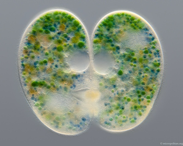
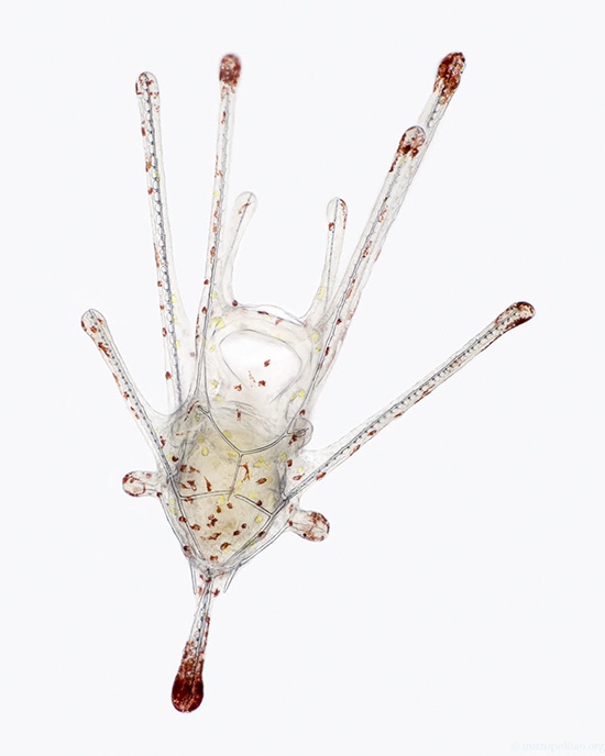
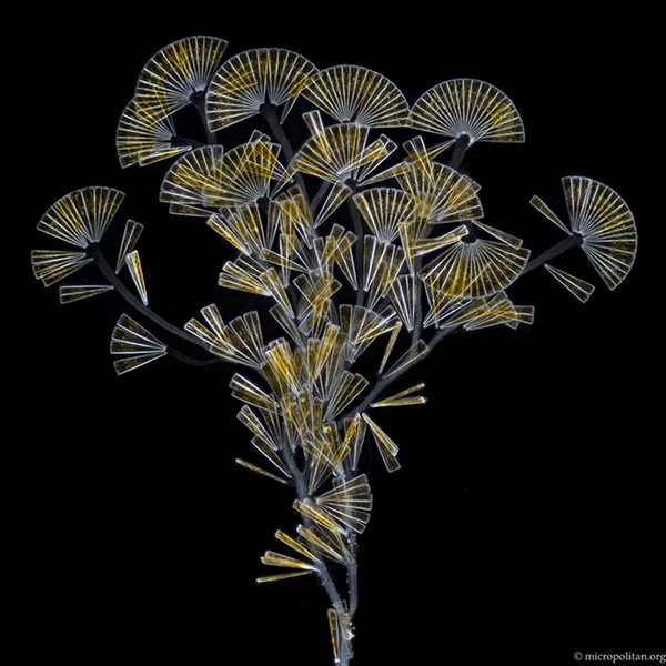
Van Egmond’s work sits at the fascinating intersection of art and science, where reality and abstraction blur. “I started as an autonomous artist, but my work was always inspired by scientific imaging,” he notes. “I was intrigued by the artificial way of depicting nature in scientific imaging.” With microscopy, he found a perfect medium to combine his artistic vision with scientific curiosity. “I try to be a realist, showing the organisms as naturalistic as possible,” he says. “But I also realize that what we see through the microscope is a projection, leaving room for interpretation.” The trick is to observe the organism meticulously and interpret its actual shape. Then try to make an image that gives a good natural impression.
His unique approach has allowed him to showcase his work in both art and science contexts. “I like to keep the question of whether what I do is art or science open-ended,” he says with a smile. “It’s fun to be able to show my work in different contexts, each with its own perspective.” Van Egmond’s images are a masterclass in balance and tranquility, inviting the viewer into a serene microscopic realm. “I like to make images that are visually convincing, even without knowing what they depict,” he explains. “Life is already chaotic enough, so it helps to have something orderly and tranquil to look at.” This approach not only creates a sense of calm but also nods to the artificial style of scientific imaging.
Specialized Techniques: Stereoscopy, Microscopy, and Time-Lapse
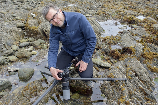
His expertise in photography through the microscope and stereoscopy has allowed him to capture the microscopic world in breathtaking detail. “Stereoscopy and microscopy are a great combination,” he notes. “When I started with microscopy, I combined the techniques, and it’s been a perfect union ever since.” He recommends starting with a stereomicroscope, which offers a stereoscopic image and is ideal for observing small objects without the need for preparations.
Filming and time-lapse movies have become essential tools in his arsenal, enabling him to convey the dynamic nature of the microscopic world. His work on the 3D IMAX film “Fungi: Web of Life” showcased his ability to create stunning 3D footage, while his time-lapse movies of fungi growth reveal the hidden lives of these organisms. By combining techniques like flash photography and macro lenses, Van Egmond provides a unique glimpse into the world beneath our feet. By using a flash photography and macro lenses in camera set up with soil behind glass plates, Van Van Egmond provides a unique glimpse into the world beneath our feet.
Historical Influences and Modern Innovations
Van Egmond’s work is deeply rooted in the history of microscopy, and he finds inspiration in the pioneering work of Antoni van Leeuwenhoek. Growing up near Van Leeuwenhoek’s hometown and now working in Delft, Van Egmond feels a strong connection to the father of microscopy. “I have always been interested in history and the early days of microscopy,” he says. “Reading Van Leeuwenhoek’s discoveries is fascinating, especially with my experience with microorganisms.”
His work is also influenced by Victorian traditions of microscopic slide making and 19th-century naturalists. He combines old-fashioned techniques with modern digital imaging, creating a unique blend of traditional craftsmanship and contemporary innovation. “Microscopy is a craft,” he notes. “The preparation of samples is essential for creating a good image. I like the combination of old-fashioned technique and modern possibilities, leading to imaging that has not been done before.”
Scientific Illustrations and Advocacy
Commissioned work presents a unique challenge for Van Egmond, requiring both artistic and scientific precision. “Microorganisms never do what you want them to do,” he laughs. “But I do commissioned work occasionally, and it’s fun when it works out well.” His images and films have been used as scientific illustrations, including a project with a mycologist friend who specializes in electron microscopy. This collaboration showcases his ability to balance art and science, creating visually stunning and informative images.
Van Egmond’s passion for promoting microorganisms stems from a desire to shift human-centric perspectives and foster appreciation for the vital role microorganisms play in our ecosystem. By sharing his captivating images, he aims to educate and inspire, dispelling fears and misconceptions surrounding microbes. “Life becomes easier when you are well-informed,” he emphasizes, encouraging self-training and exploration.
Contributions to Micscape Magazine and the Micropolitan Museum
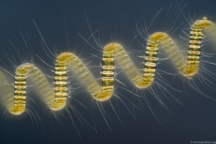
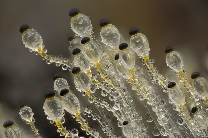
Van Egmond’s contributions to Micscape Magazine and the online Micropolitan Museum have been instrumental in promoting the study of microorganisms. Although he hasn’t contributed to Micscape recently due to workload, his past articles aimed to inspire beginners in microscopy. The Micropolitan Museum, created over 20 years ago, showcases his micrographs in a lighthearted and accessible way, making it an excellent introduction for newcomers to the field.
Unveiling the Secrets of Antoni van Leeuwenhoek’s Discovery
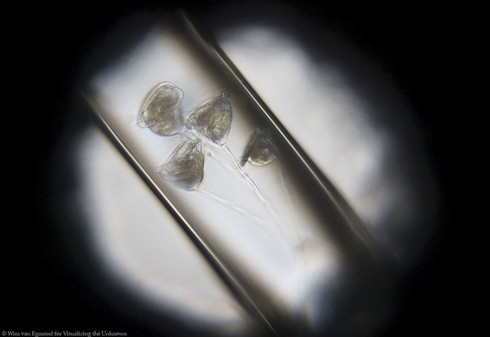
Van Egmond’s current research focuses on Antoni van Leeuwenhoek’s first discovery of microorganisms. Living at the exact spot where Leeuwenhoek made his groundbreaking find, Van Egmond has delved into the history, arguing that Leeuwenhoek discovered a cyanobacteria bloom. This research has led to collaborations with historians and involvement in the “Visualizing the Unknown” project, which explores the intersection of art, science, and history.
Upcoming Projects and Future of Microscopy and Photography
Van Egmond is excited about future projects combining his imagery with music, a long-standing passion. He collaborates with musicians and composers, creating immersive experiences that showcase the beauty of microorganisms. With ongoing research and creative projects, Van Egmond continues to inspire and educate, promoting a deeper appreciation for the microscopic world.
He envisions a future where microscopy and photography continue to evolve, driven by technological advancements and artistic innovation. Van Egmond predicts that techniques like focus stacking and artificial intelligence will become increasingly accessible, enabling the creation of breathtaking images that blend art and science. However, he also emphasizes the importance of the personal touch and imperfections that make an image truly unique.
Advice for Aspiring Photographers and Scientists
Van Egmond offers valuable advice to those interested in exploring the microscopic world: be patient, take your time to master the microscope, and connect with others who share your passion. Joining microscopy clubs or online forums can provide valuable resources and inspiration.
Personal Reflections and Long-term Goals
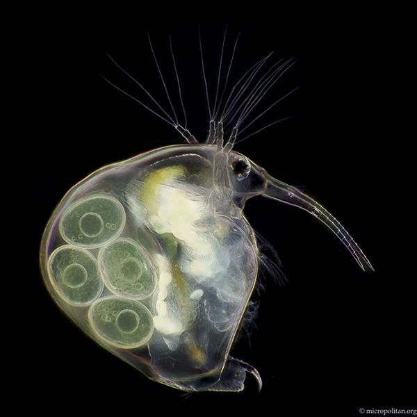
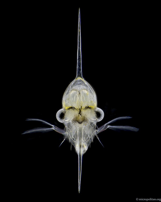
Looking back on his career, Van Egmond is proud of his achievements, including working with Leeuwenhoek’s microscopes, helping create Micropia, and contributing to films like Fantastic Fungi. However, he emphasizes that the true joy lies in the work itself, not just the recognition. He finds inspiration in collaboration, community, and the thrill of discovery. If he had to choose his own favourite piece of work it would be one with humor. Like the movie of a water flea playing the piano, which can be viewed here.
His long-term goals include continuing to explore the microscopic world, collaborating with others, and perhaps revisiting his early passion for drawing microorganisms. He remains driven by curiosity and the pleasure of creating, always seeking new ways to inspire and educate others about the wonders of the microscopic realm.
Inspiring the Next Generation and Lasting Impact
Van Egmond hopes that his work will inspire people, especially children, to explore the micro-world themselves. He recently developed a course for kids in the Netherlands, where they learned about science through the eyes of Leeuwenhoek, even making their own microscopes using simple DIY methods. He believes that sparking interest in natural history at a young age can lead to a lifelong passion for discovery. Van Egmond encourages everyone to explore the micro-world, emphasizing that microscopes don’t have to be expensive, and even a good hand lens or smartphone can reveal the amazing world of microbes.
He has received heartfelt feedback from scientists who have seen their subject of study in a new light through his footage. Some have even become emotional, having never seen their area of research presented in such a unique and captivating way. These reactions remind Van Egmond of the power of his work to inspire and educate, leaving a lasting impact on his audience.