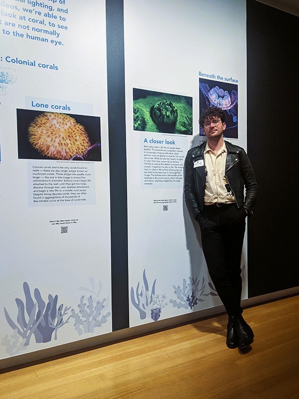
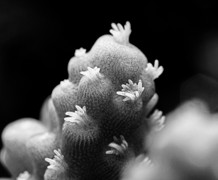
What are coral? - DLSR macro image of a coral branch and its constituent polyps in Acropora sp.. Corals are colonial organisms made up of anywhere between 1 and 1 million polyps all interconnected with each other by a layer of tissue. This allows them to communicate with each other and share nutrients and proteins quickly across the colony when needed. The black dots in this image are the symbiotic algae cells that reside in the coral tissue and photosynthesise providing the coral with nutrients., Courtesy: Brett Lewis
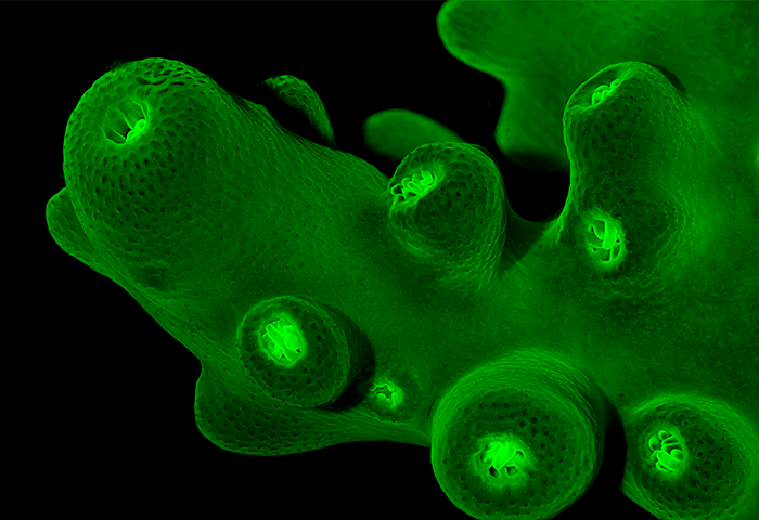
Coral branches glow under moonlight - Stereo micrograph of the green fluorescent proteins in the branching coral Acropora sp. When exposed to blue wavelengths of light the proteins in the coral tissue 'glow' giving researchers and understanding of how healthy the colony is., Courtesy: Brett Lewis
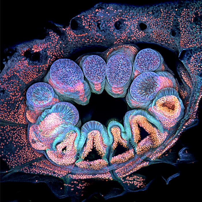
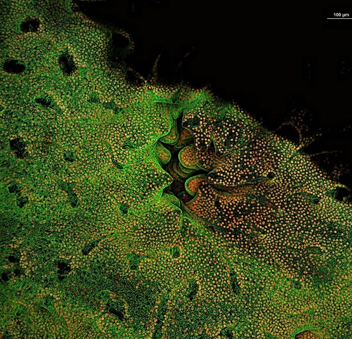
Autofluorescence micrograph of the polyp of reef-building coral Pocillopora verrucosa - Nikon Small World 2022 winner. An inverted confocal fluorescence micrograph of a polyp in the coral Pocillopora verrucosa. The polyp is the animal, of which there can be thousands, that makes up the coral colony. The colours in this image represent proteins fund in the cells of the coral polyp, giving researcher's insight into cell function and colony health., Courtesy: Brett Lewis
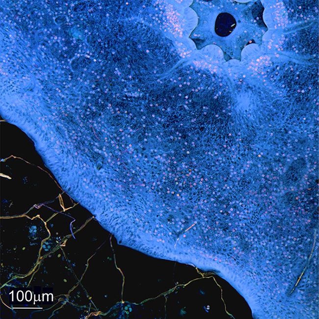
Autofluorescence micrograph of the polyp of reef-building coral Montipora hispida. - Inverted confocal fluorescence micrograph of a polyp in reef-building coral Montipora hispida which has more green fluorescent proteins than P. verrucosa. The polyp in this coral in M. hispida is also smaller and has retracted back into the safety of the corallite, which it does if it feels threatened., Courtesy: Brett Lewis
Top down look at the coral tissue as it grows over the coral reef forming a lasting bond that can survive waves forces created by cyclones and tsunamis., Courtesy: Brett Lewis
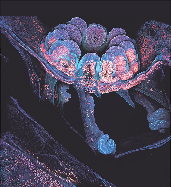
Looking inside the coral; Autofluorescence cross-sectional micrograph of the polyp of reef-building coral P. verrucosa. This image is a via into the coral colony and the dark passageways that transfer nutrients across the colony. The spiralling structures are the digestive mesenterial filaments which wrap around and digest any prey that is unlucky enough to get capture by the polyp. The long-striated tissue is the polyp muscles which help retract the polyp when it is in danger., Courtesy: Brett Lewis