Montipora Polyp Fluro Micro - Inverted confocal fluorescence micrograph of a polyp in reef-building coral Montipora hispida which has more green fluorescent proteins than P. verrucosa. The polyp in this coral in M. hispida is also smaller and has retracted back into the safety of the corallite, which it does if it feels threatened by Brett Lewis, Marine biologist, Micrographist, Australia

The Beauty of the Coral Polyp By Brett Lewis, ​One of the Winners of the Nikon Small World 2022 Photomicrography Competition

Self portrait by Brett Lewis, Marine biologist, Micrographist, Australia
Self portrait, Courtesy: Brett Lewis

The Beauty of the Coral Polyp By Brett Lewis, ​One of the Winners of the Nikon Small World 2022 Photomicrography Competition

Brett Lewis has an avid passion for corals and producing visual science. He has dedicated himself to documenting and researching corals around the coasts of Australia. Brett utilizes advanced microscopic technology when taking images of coral tissues and cells to gain further knowledge about the organisms and their behaviors. The images of corals taken by Brett are highly acclaimed due to their detailed and clear resolution. His imagery, research, and videos were used in the Reef Rescue documentary which “unveils the groundbreaking science behind ‘super corals’ and a radical new plan to save the Earth’s imperiled coral reefs” – has won awards on Netflix. In fact, Brett’s work features in 10+ documentaries and over 30 museums and ocean education centers globally.

Recently, his image of the autofluorescence of a single coral polyp won the top 20 of the prestigious Nikon Small World 2022 Photomicrography Competition. In the following interview, you will get to know Brett and his love for corals.

DLSR macro image of a coral branch and its constituent polyps in Acropora sp.. The black dots are the symbiotic algae cells that reside in the coral tissue and photosynthesise to give the coral nutrients. by Brett Lewis, Marine biologist, Micrographist, Australia
What are coral? - DLSR macro image of a coral branch and its constituent polyps in Acropora sp.. Corals are colonial organisms made up of anywhere between 1 and 1 million polyps all interconnected with each other by a layer of tissue. This allows them to communicate with each other and share nutrients and proteins quickly across the colony when needed. The black dots in this image are the symbiotic algae cells that reside in the coral tissue and photosynthesise providing the coral with nutrients., Courtesy: Brett Lewis

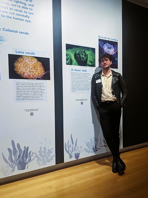
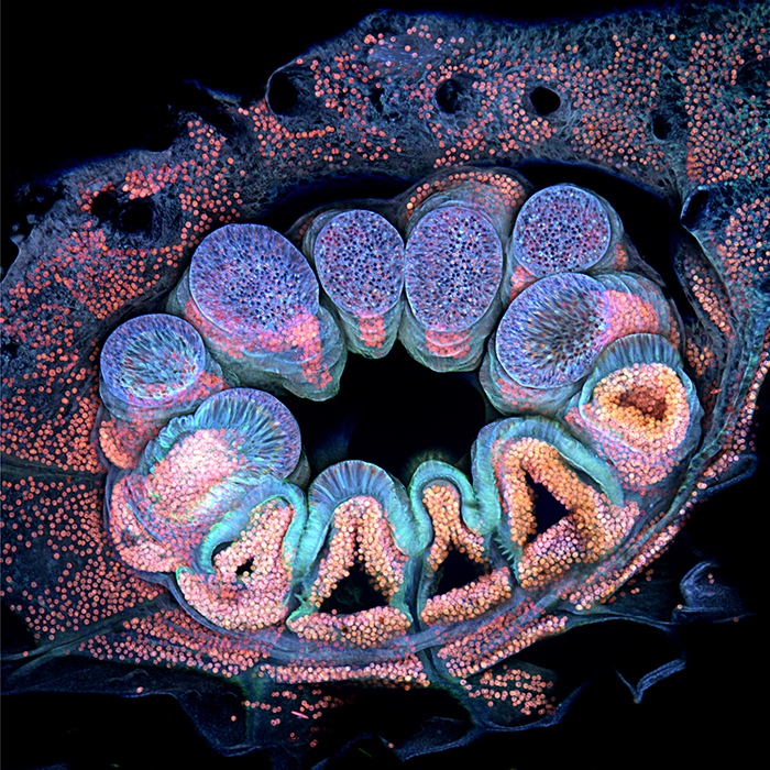
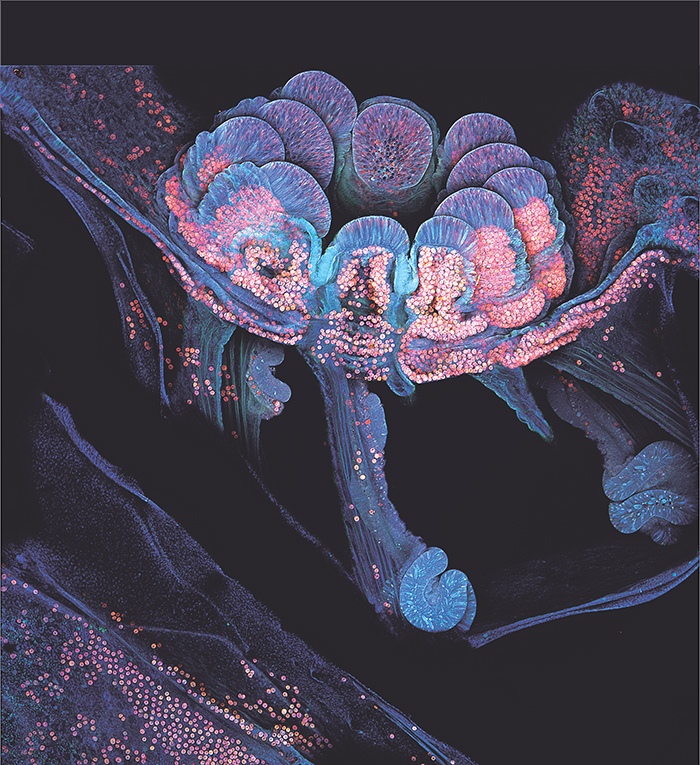
Q: Congratulations on your image of the Autofluorescence of a single coral polyp winning the top 20 of the prestigious Nikon Small World 2022 Photomicrography Competition. Please tell us about your background and training in becoming an outstanding micrographist.
A: An outstanding micrographist is very flattering, thank you!

Just like everything in life, becoming a microscopist and imaging coral really wasn’t a part of the plan – I wanted to be Dr. Alan Grant from the film Jurassic Park, haha.

That changed when I started travelling Australia in my early 20s and spent time on the Great Barrier Reef, Australia, working on tropical islands and spending a lot of time underwater exploring the reef. I fell in love with the reef’s beauty, its colors, and its life. Here, I started to learn photography, to capture the reef’s remarkable environments, and ended up working in a large outdoor aquarium on Daydream Island where I would give tours, educate visitors, and look after the inhabitants such as sharks and stingrays.

It was here that I decided I wanted to become a marine scientist.

At first, I wanted to continue working with sharks and stingrays but there wasn’t much research in this area at my university. So, I ended up working with something much smaller.

In my first year of university, I was asked to do some research assistant work looking at coral fossils – not dinosaurs but it’ll do. So, in an attempt to make the most out of this very cool opportunity I brought my photography skills to this task, using any photographic equipment I could find including some simple microscopes. Anything to impress and hopefully get my supervisors to keep me on.

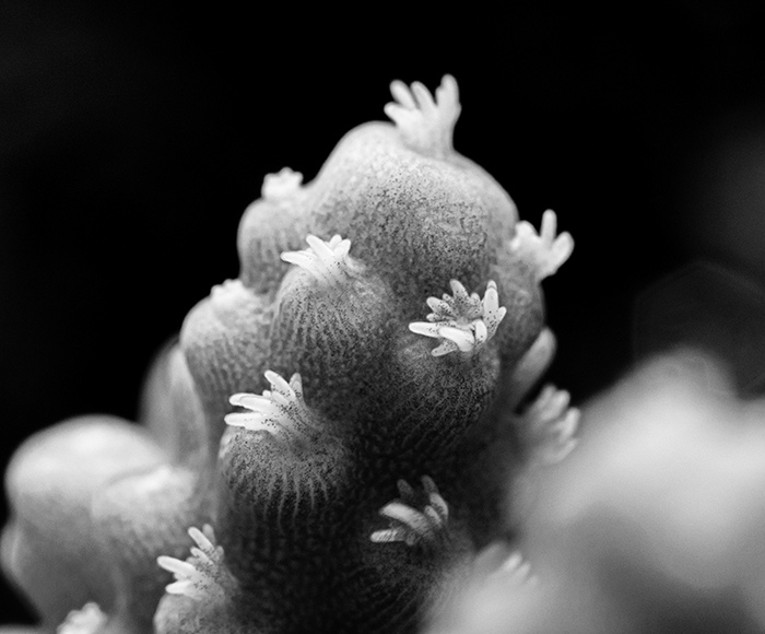
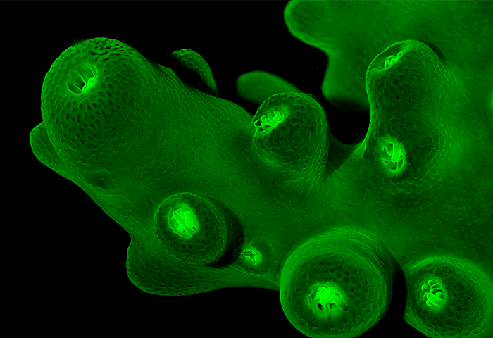
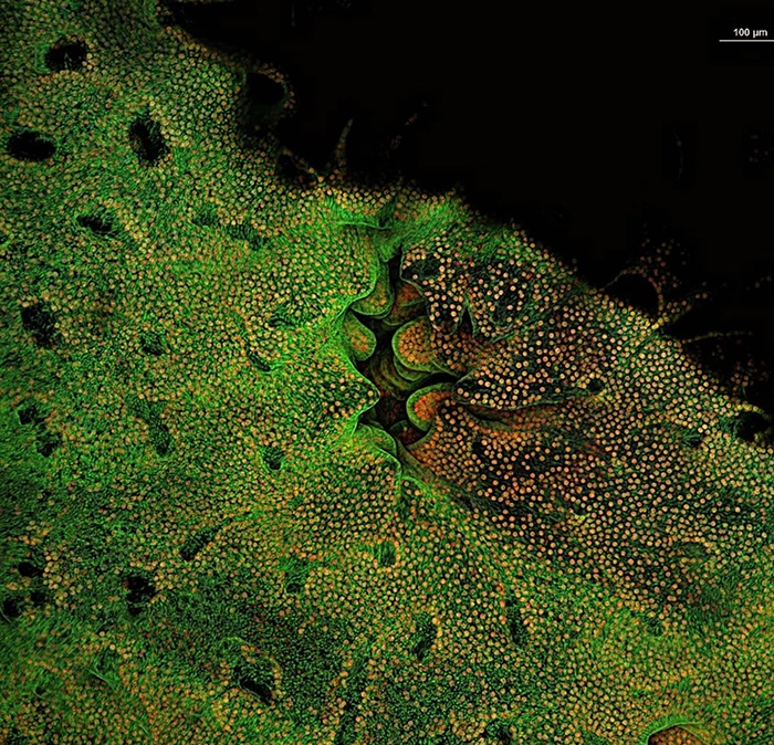
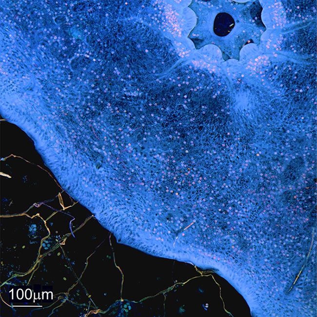
It worked, I have now completed my Masters’s degree where I modernized techniques to create time-resolved high-resolution imagery of coral behavior, cells, and subcellular structures that help us understand how corals adapt the external pressures and stress. By the end of 2022, I will have submitted my Ph.D. in which I have further modernized microscopy techniques in coral by incorporating the coral’s natural autofluorescence with improved resolution and developed techniques to render the cells and subcellular structures of a coral in three dimensions.

Stereo micrograph of the green fluorescent proteins in the branching coral Acropora sp.by Brett Lewis, Marine biologist, Micrographist, Australia
Coral branches glow under moonlight - Stereo micrograph of the green fluorescent proteins in the branching coral Acropora sp. When exposed to blue wavelengths of light the proteins in the coral tissue 'glow' giving researchers and understanding of how healthy the colony is., Courtesy: Brett Lewis

Q: Please share with us the story behind this winning image.
A: This image was an attempt to push the novel imaging techniques to their fullest. I had observed a few nice images of coral polyps but I felt there was an opportunity to produce even more detailed images – plus I wanted to have some fun!

This image is an individual coral polyp (1-3mm) sitting in the coral colony’s interlocking tissue.
The polyp was imaged using 4 scanning lasers with very specific colors.

I took the image over 36 hours which is anywhere from 70 to 100 images stacked together (z-stacked) and can be rendered in 3D. Each image takes 20 to 30 minutes to produce.

By imaging the autofluorescence of the coral we can identify a fluorescence emission signature for the tissues and observe when and where these processes shift, including the individual cells. These changes can be mapped and plotted to be compared throughout time and across coral species, helping to identify when a colony is healthy and when it may be under stress or going through change.

The pink/orange cells are the coral’s photosynthetic zooxanthellae; the algae cells that live within the coral’s tissue and provide the colony with much of its nutrients.

The bulbous ends of the polyps tentacles are filled with stinging cells for capturing and paralyzing prey (red circles, black voids, and purple hue). The cyan coloration is primarily from the coral’s simple supporting cells and the mucus-producing cells.

Montipora Polyp Fluro Micro - Inverted confocal fluorescence micrograph of a polyp in reef-building coral Montipora hispida which has more green fluorescent proteins than P. verrucosa. The polyp in this coral in M. hispida is also smaller and has retracted back into the safety of the corallite, which it does if it feels threatened by Brett Lewis, Marine biologist, Micrographist, Australia
Autofluorescence micrograph of the polyp of reef-building coral Pocillopora verrucosa - Nikon Small World 2022 winner. An inverted confocal fluorescence micrograph of a polyp in the coral Pocillopora verrucosa. The polyp is the animal, of which there can be thousands, that makes up the coral colony. The colours in this image represent proteins fund in the cells of the coral polyp, giving researcher's insight into cell function and colony health., Courtesy: Brett Lewis

Q: What is it about coral colonies that fascinate you the most? How many types/families of corals are on our planet earth?
A: There are about 600+ species on Earth. But who knows if there are other coral-like creatures elsewhere in the universe? They’re simple organisms whose lineage appeared during the Palaeozoic some 250 million years ago. If multicellular ocean life exists elsewhere in the universe, it may look like coral.

What fascinates me… that corals are efficient hunters even though most can’t move.

Coral polyps possess stinging cells in their tentacles to subdue prey and drag it into its mouth, which you can see in this image.

Inside the mouth is the stomach but within the stomach are long digestive filaments that encompass and devour the prey. These digestive filaments can be extended outside of the coral body through the tissues in defense against competitors and other coral colonies.

Now imagine if polyps were larger, it would be a worse Sarlacc pit.

The natural colours of a Trachaphyllia coral polyp who possess ancient structures that date back to corals 250 million+ years ago and are not present in 'modern' reef-building coral. We are now investigating this species to garner insights into coral adaptive evolution to see how modern coral evolved. by Brett Lewis, Marine biologist, Micrographist, Australia
Autofluorescence micrograph of the polyp of reef-building coral Montipora hispida. - Inverted confocal fluorescence micrograph of a polyp in reef-building coral Montipora hispida which has more green fluorescent proteins than P. verrucosa. The polyp in this coral in M. hispida is also smaller and has retracted back into the safety of the corallite, which it does if it feels threatened., Courtesy: Brett Lewis

Q: In your opinion, how severe is the coral bleaching event? Are there any solutions for this event currently?
A: Bleaching is a huge concern, but it is only the symptom, a sickness, a breakdown of the coral’s relationship with its symbiotic algae (zooxanthellae) that creates a toxic feedback loop in the coral as the algae begin to oxidize. The coral must expel the algae to remove the toxicity. Unfortunately, that also removes the corals primary major source of energy. Sometimes corals will recover if conditions return to normal quickly. However, these periods of high temperatures are becoming longer as the planet’s climate changes and the amount of time for the coral to recover between events is becoming less. As a result, the bleaching and coral reef die-offs will worsen unless we curb emissions and the only way to curb emissions is to stay informed, put pressure on politicians, and vote for change.

Top down look at the coral tissue as it grows over the coral reef forming a lasting bond that can survive waves forces created by cyclones and tsunamis. By Brett Lewis, Marine biologist, Micrographist, Australia
Top down look at the coral tissue as it grows over the coral reef forming a lasting bond that can survive waves forces created by cyclones and tsunamis., Courtesy: Brett Lewis

Q: What equipment and technology did you use to take such crisp, clear images of marine life?
A: Corals were fixed, dehydrated, and set into specialized imaging resin. The resin was cut and polished to expose the imaging surface. A process that takes up to three weeks per sample.
The surface was imaged using the Nikon A1R inverted confocal laser scanning microscope. The laser bandwidths used were 360 nm (Blue) 488 nm (Green), 546 nm (Red), and 655 nm (Near-infrared) with a dwell time of 20 to 30 minutes.

Corals were housed in research aquariums after being collected off the Great Barrier Reef and transported to the research systems at Queensland University of Technology (QUT).

Looking inside the coral; Autofluorescence cross-sectional micrograph of the polyp of reef-building coral P. verrucosa. This image is a via into the coral colony and the dark passageways that transfer nutrients across the colony. The spiralling structures are the digestive mesenterial filaments which wrap around and digest any prey that is unlucky enough to get capture by the polyp. The long-striated tissue is the polyp muscles which help retract the polyp when it is in danger., By Brett Lewis, Marine biologist, Micrographist, Australia
Looking inside the coral; Autofluorescence cross-sectional micrograph of the polyp of reef-building coral P. verrucosa. This image is a via into the coral colony and the dark passageways that transfer nutrients across the colony. The spiralling structures are the digestive mesenterial filaments which wrap around and digest any prey that is unlucky enough to get capture by the polyp. The long-striated tissue is the polyp muscles which help retract the polyp when it is in danger., Courtesy: Brett Lewis

Q: You are also working with museums in their installations. Please tell us more about this line of work and give us some examples of your accomplishments.
A: Our work has been featured in over 30 national and international museums and aquariums over the last four years.

This has led to my work winning or being nominated for 12 international and national awards.

Currently, we have an exhibit at QUT Art Museum where we used AR to bring coral to life as part of a collaboration with Japanese artists Julia and Ken Yonetani.

The media produced from our research has been included in 13 separate documentaries, including the award-winning documentary Reef Rescue on Netflix.

I am also a councillor for the Australian Coral Reef Society (ACRS) focused on coral reef research.
As a celebration of 100 years of coral reef sciences, we at the ACRS have put together a coffee table book and museum exhibit at the Queensland Museum documenting the rich history of coral reef science in Australia.

Q: What is your advice for the general public regarding helping the coral reefs?
A: Listen to the experts, be critical and vote in favor of politicians and policies which can help curb emissions.

The first ever look at coral cells in 3 dimensions (3D). This is a 3D render of 100 focused ion beam scanning electron micrographs, capturing the corals cells ultrastructures (what makes up a cell). In this case we are looking at a storage cell full of lipid vesicles (purple), the cell nuclei blue, cilia and microvilli (long and short hairs on the top of the cell) and the cell membrane (pink). By Brett Lewis, Marine biologist, Micrographist, Australia

*****

We thank Brett for sharing his award-winning image and his knowledge of corals. We hope his advocacy for corals inspires many more people to protect the reefs.

Brett’s Instagram