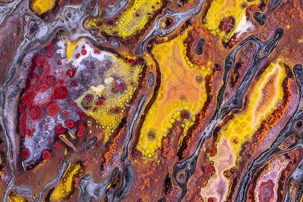
It was found in the Morrison Formation, where dinosaur remains are prevalent. The age of this geologic formation corresponds to that of the middle part of the dinosaur period, the Jurassic, when there were no other very large animals. Finally, the bone itself is five or six times the thickness of the thickest long bone in an adult male human and, therefore, it had to have come from a very large beast indeed. Contrary to all appearances, the red structure within the quartz-filled medullary cavity is not a nutrient artery. Soft tissue rarely becomes fossilized, and when it does, it is rarely replaced by quartz. The red color comes principally from hematite, an iron oxide. The bone in the lower right is metaphyseal bone, also called spongy bone, with blue chalcedony between the spicules. Courtesy: Norm Barker
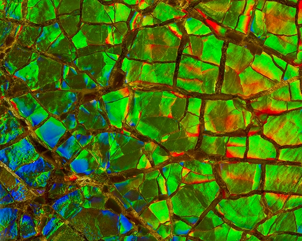
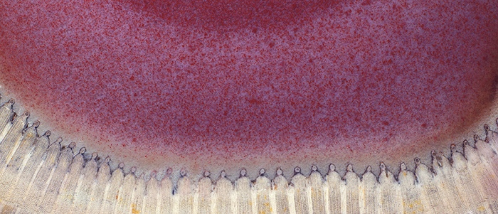
This photograph shows ammonite shell under high magnification. Specimens such as this are sold for jewelry under the name amolite. Courtesy: Norm Barker
二十年前,我与友人合著了一本书,名为《远古微观世界》,”Ancient Microworlds”,该书在过去的十二年中一直在美国、欧洲和亚洲,做巡回展出。超过一百万的博物馆参观者看过这本书中所登载的图像。我的作品也曾在史密森学会、纽约自然历史博物馆、伦敦科学博物馆以及堪萨斯城的纳尔逊-阿特金斯艺术博物馆等场所展出。我的个人作品被40多家博物馆永久收藏。另一本名为《海藻:海洋奇观》,“Seaweeds: Wonders of the Ocean Realm”的书也有巡回展览,并在2006年,开幕于南卡罗来纳州的查尔斯顿博物馆。
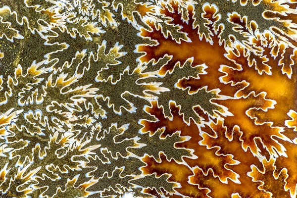
This early fossil ammonite shows the suture marks that have formed on the animals outside shell. Suture patterns of an ammonite Placenticeras intercalare. The inside of the shell is divided into a chambered part and an unchambered part. The body of the animal occupied the unchambered part, or living chamber. The chambered portion of the shell was divided by partitions called septa. The contact of these septa with the external shell is called a suture.
It is from the Late Cretaceous period and from The Bear Paw Formation, McGrath, Alberta Canada.
The ammonite lived throughout the Jurassic and Cretaceous periods of history, 146–203 million years ago. Courtesy: Norm Barker
问: 请与我们分享你最近出版的书《隐藏之美:探索医学中的美学”》,“Hidden Beauty: Exploring the Aesthetics of Medical Science”。是什么促使你写这本书? 答: 显然,我们在研究中看到了令人著迷的人体,人体是一台了不起的天然机器。我们也喜欢接触科学中所有不同的知识分支。有一天,我可能会拍摄一头大象的尸检,它的心脏看起来几乎像人类的心脏,但重达40磅,或者我可能会拍摄稀有的15世纪手稿。我们永远不知道大型学术医疗中心会要求我们承担什么样的任务。Shiffer图书出版的《隐藏的美丽:探索医学美学》一书的整个想法是将艺术和科学相结,展示了艺术家和科学家的合作成果。我们想分享在研究时所观察到的人体之美,其中使用了MRI,电脑和显微镜等,所有不同类型的成像方式,
Arthropitys bistriata, x10 Permian Araguaina, Brazil Modern plants of the calamities family are the horsetails of which this specimen is a species. The white in this photograph is the xylem, or true wood, and the pink is the pith or center of the stem, which has been filled with quartz colored pink by iron. Because a tube is a structure that resists bending, calamities of relatively narrow diameter could grow into plants that were more than a hundred feet tall. Courtesy: Norm Barker
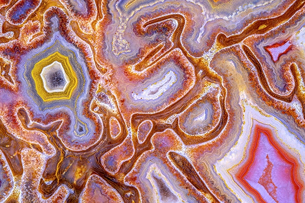
Quartz and some of the associated minerals that give it color enter into the fossilizing process as solutions, gels, and sols. Although they move as liquids, gels and sols are made up of very small particles bound together by weak electrical bonds. In this specimen, the linear arrays of small round structures were produced by local condensations of particulate mineral. During their consolidation, small puddles of pigment formed that are unrelated to the structure of the bone itself. Courtesy: Norm Barker
问: 根据你的专业经验,你从人体和自然中学到了什么? 答: 人体是一台极其复杂的机器,即使采用了所有的21世纪技术,仍然不知道还有多少令人惊讶的未知事物,等待著我们发掘,例如,人类的大脑。就在100多年前,神经外科这个亚专业出现了,这是因为西班牙病理学家和显微镜学家Santiago Ramón y Cajal发现了大脑通过中细胞突触之间发送信号,进行交流。他的这项发现,引发了一场革命,他也因此获得了诺贝尔奖,并被认为是现代神经科学之父。他还是一位艺术家,他用显微镜绘制了自己的观察插图,这些插图本身就是令人惊叹的美丽艺术品。进入2022年,令人惊讶的是,虽然我们对这个复杂的器官已经了解了许多,但未知的领域还有很多,我们将继续研究和学习。
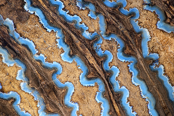
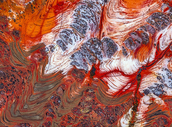
Biwabik Iron Formation stromatolites, made up of swirls of predominantly red and metallic hematite, bear witness to the previous presence of cyanobacteria 3.5 billion years ago. For at least half the time that life has existed on earth, bacteria were the only forms of life and, during this time, those with blue-green pigment that could photosynthesize (cyanobacteria) significantly changed the atmosphere by adding oxygen. Originally the earth's atmosphere was like that of Mars and Venus, with probably less than one percent oxygen. Photosynthetic cyanobacteria produced not only oxygen from carbon dioxide but also storable high-energy chemical compounds like adenosine triphosphate. These compounds permitted higher forms of life to evolve by allowing locomotion and other physiologic activities to develop. Courtesy: Norm Barker