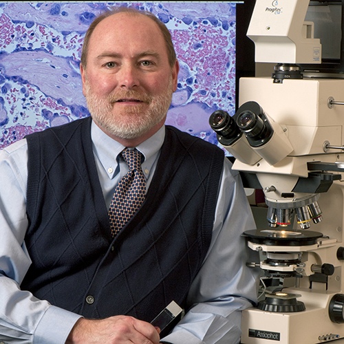
We are honored to be able to speak with Professor Norm Barker who is the Director of the Pathology Photography and Graphic Arts Laboratory at the Medical School of Johns Hopkins University. Professor Barker has a keen eye for beauty in nature under his microscope. The images captured by him span a wide range, from billion-year-old fossils to living cells inside a human body. His photographs have illustrated more than fifty textbooks and he has published numerous photo essays in major magazines. His work is in the permanent collections of more than forty museums including The Smithsonian, The George Eastman House, The American Museum of Natural History, the Nelson-Atkins Museum, and the Science Museum in London.
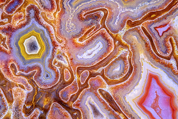
It was found in the Morrison Formation, where dinosaur remains are prevalent. The age of this geologic formation corresponds to that of the middle part of the dinosaur period, the Jurassic, when there were no other very large animals. Finally, the bone itself is five or six times the thickness of the thickest long bone in an adult male human and, therefore, it had to have come from a very large beast indeed. Contrary to all appearances, the red structure within the quartz-filled medullary cavity is not a nutrient artery. Soft tissue rarely becomes fossilized, and when it does, it is rarely replaced by quartz. The red color comes principally from hematite, an iron oxide. The bone in the lower right is metaphyseal bone, also called spongy bone, with blue chalcedony between the spicules. Courtesy: Norm Barker
His recent image, “Agatized dinosaur bone”, was awarded Image of Distinction by the 2021 Nikon Small World Photomicrography Competition. It was derived by taking a small fragment of a dinosaur bone dated to 1 billion years ago. Thanks to micographists like Norm, images of rare fossils come to life, viewable by the public. Also, by connecting art with science, micrographists help people appreciate nature in a way that they would not ordinarily experience.
We hope you enjoy the following interview.
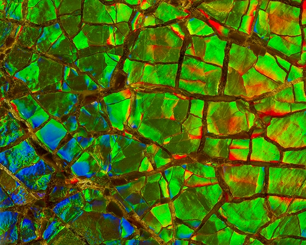
This photograph shows ammonite shell under high magnification. Specimens such as this are sold for jewelry under the name amolite. Courtesy: Norm Barker
Q: Please share with us your path in becoming an expert in photomicroscopy and macro photography. A: After I graduated from The Maryland Institute College of Art with a BFA in Photography, I wanted to go on to graduate school but had to pay the bills. I fell into a two-year paid training position at the Johns Hopkins School of Medicine as a biomedical photographer. At the time I didn’t even know what a biomedical photographer was, I wanted to be an artist. It didn’t take long to figure that art was not going to pay the bills. This was in the late 70s and one of the benefits of the job was that they paid for graduate school in the evening as long as you went to Johns Hopkins. I completed an M.S. in Education as well as an M.A. in Publications Design, while working at Hopkins during the day and after I finished my training was hired on staff.
As a biomedical/scientific photographer and tenured full Professor of Pathology and Art as Applied to Medicine at Johns Hopkins University in Baltimore, Maryland, I serve as director of the Pathology Photography & Graphic Arts Laboratory. My work has been published in magazines, books, and exhibited around the world in museums and traveling exhibitions. My work emphasizes the incredible diversity of the photographic community; armed with a microscope and other, more conventional, photographic equipment, I bring people to a world not seen with the human eye.
Agatized Dinosaur Bone, Courtesy: Norm Barker
Q: Your “Agatized dinosaur bone” won the Image of Distinction award in the 2021 Nikon Small World Photomicrography Competition. Congratulations! Please share with us the story of this image and what equipment you used in capturing the image. A: I’ve been at Hopkins for 41 years and I can honestly say it’s been a little of everything. The unusual thing about a biomedical/scientific photographer is that he or she needs to be an expert with all the specialized techniques used for imaging science. But they also need to be highly proficient with all kinds of different areas of imaging such as portraiture, studio/location, public relations, architecture, just to mention a few. These are areas that we cover on any given day as our normal job responsibilities. Working in a pathology department I specialize in photomicrography, (photography under the microscope). The microscope provides a whole different beautiful world, that if done correctly can provide an interface between both science and art. It’s amazing when you hold some of these specimens in your hand and you realize that in many cases the fossilized material is hundreds of millions of years old!
This small piece of dinosaur bone was photographed with a Zeiss microscope and a special Zeiss Luminar, f4.5, 40 mm macro lens with an RMS thread. The lighting was done with fiber optics and the specimen was imaged with being coated with mineral oil to reduce any reflections and cut marks. Over millions of years the actual specimen has been mineralized or replaced with silica compounds such as agate, jasper, opal or chalcedony.
Gomphothere Tooth, Courtesy: Norm Barker
Q: You seem to have a deep interest in Ancient Microworlds. Please share with us some of your findings and your advice on the techniques required in photographing fossil slabs. A: One of the great things about my job is that I get to collaborate with some of the top scientists and researchers in the world. I’ve had work published in everything from scientific images in Nature to portraits in Penthouse, Smithsonian, and Natural History. I have also had the pleasure of working on more than 50 textbooks, fascicles, and atlases, where I did all the photography and have provided thousands of photomicrographs for the books. I just published a chapter about Photography of Fossils, Minerals and Geological Photography, in a book called “Natural Science Imaging and Photography”, Edited by Michael Peres, and published by Focal Press.
Twenty years ago I co-authored a book, titled “Ancient Microworlds”, which has an exhibit that has been traveling in the U.S., Europe, and Asia for the last twelve years. More than a million museum visitors have seen the show.
Some of the other venues where my work has been exhibited include The Smithsonian Institution, The Museum of Natural History in New York, The Science Museum in London, as well as the Nelson-Atkins Museum of Art in Kansas City. My personal work is in the permanent collections of more than 40 museums. The other book titled “Seaweeds: Wonders of the Ocean Realm” has a traveling exhibit as well and opened at The Charleston Museum in South Carolina in 2006.
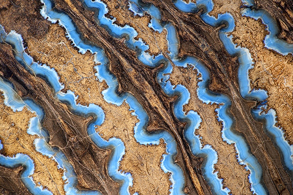
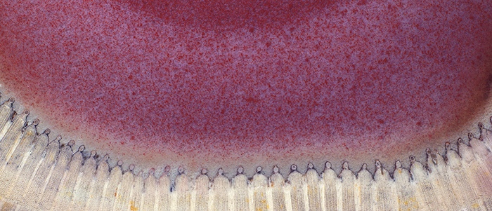
This early fossil ammonite shows the suture marks that have formed on the animals outside shell. Suture patterns of an ammonite Placenticeras intercalare. The inside of the shell is divided into a chambered part and an unchambered part. The body of the animal occupied the unchambered part, or living chamber. The chambered portion of the shell was divided by partitions called septa. The contact of these septa with the external shell is called a suture.
It is from the Late Cretaceous period and from The Bear Paw Formation, McGrath, Alberta Canada.
The ammonite lived throughout the Jurassic and Cretaceous periods of history, 146–203 million years ago. Courtesy: Norm Barker
Q: Please share with us your recent book, “Hidden Beauty: Exploring the Aesthetics of Medical Science”. What motivated you to write this book? A: Obviously we get a fascinating behind the scenes look at the human body, which is an amazing machine, but we also enjoy being exposed to all different branches of knowledge in science. One day I might photograph the necropsy of an elephant, whose heart looked almost human but weighed in at 40 pounds, or I might be photographing rare 15th-century manuscripts. We just never know what assignment we might be asked to cover for a large academic medical center. The whole idea of the book “Hidden Beauty: Exploring the Aesthetics of Medical Science”, published by Shiffer Books, was an artistic and scientific collaboration to show an artist and a scientist working together. We wanted to share that behind the scenes look at the beauty of the human body using all different types of imaging modalities with everything from MRI to computers and microscopes.
Q: What is anatomic pathology? How is anatomic pathology utilized in diagnosing diseases? How is it different from other forms of imaging? A: Anatomic Pathology is a medical specialty involved with the study and diagnosis of disease based on microscopic evaluation of tissues and organs, along with autopsy procedures. In the whole discipline of pathology there are many specialty areas, such as clinical lab medicine, surgical and forensic pathology to name a few. At a large academic medical center you have several pathologists that might just specialize in one organ system, like a Renal Pathologist, GYN Pathologist, or Neuropathologist.
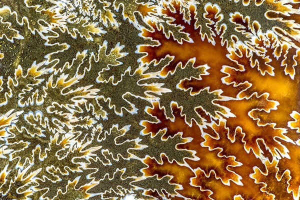
Arthropitys bistriata, x10 Permian Araguaina, Brazil Modern plants of the calamities family are the horsetails of which this specimen is a species. The white in this photograph is the xylem, or true wood, and the pink is the pith or center of the stem, which has been filled with quartz colored pink by iron. Because a tube is a structure that resists bending, calamities of relatively narrow diameter could grow into plants that were more than a hundred feet tall. Courtesy: Norm Barker
Q: Please share with us some of your memorable experiences in your profession. A: That’s a difficult question to answer; over the years I photographed a lot of very unusual stuff. But the most interesting thing about my job is I get to work with some incredibly smart and creative people. Everything from graduate students, residents, fellows, and postdocs and of course many of the top medical minds in the world. I really enjoy teaching the medical illustrators in the Department of Art as Applied to Medicine. They are incredibly talented and their work and creativity is amazing.
Q: What is your advice for aspiring micrographists especially in the field of medicine? A: A microscopist in medicine is generally associated with a particular discipline or specialty area, for example, Cell Biology or Ophthalmology or Pathology. They use the microscope as a tool for discovery and for documentation and publication of the images used for research in scientific journals or textbooks.
There are many amateur microscopist’s out there who use the microscope as a way of exploring an unseen world. Most people’s first encounter with a microscope is in high school biology. In the 1800s and turn of the century, the microscope was used by many as a tool for entertainment and discovery into the micro world. From there some go on to be inspired to study science or medicine.
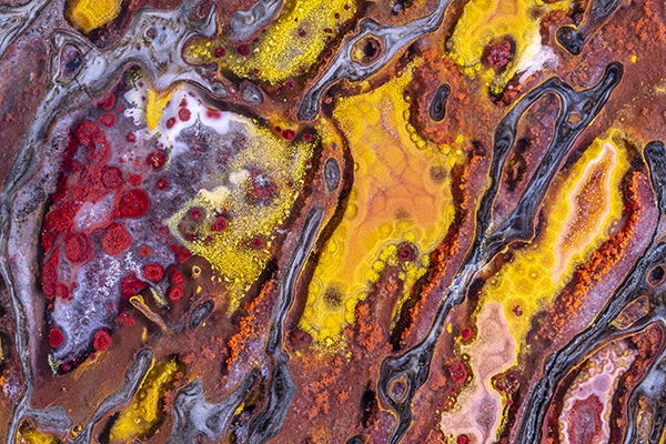
Quartz and some of the associated minerals that give it color enter into the fossilizing process as solutions, gels, and sols. Although they move as liquids, gels and sols are made up of very small particles bound together by weak electrical bonds. In this specimen, the linear arrays of small round structures were produced by local condensations of particulate mineral. During their consolidation, small puddles of pigment formed that are unrelated to the structure of the bone itself. Courtesy: Norm Barker
Q: Based on your professional experience, what are some of the lessons that you have learned from human bodies and nature? A: The human body is an incredibly complex machine and even with all of our 21st-century technology, it’s amazing how much we don’t know. For example, when you take a look at the human brain, it was just over 100 years ago when the subspecialty of Neurosurgery took place and that started a revolution with the Spanish pathologist and microscopist Santiago Ramón y Cajal figuring out that the brain communicated through electric signals sent between cell synapses. He won a Nobel Prize for his work and is considered the father of modern neuroscience. He was also an artist and drew his own illustrations of observations with the microscope which are incredibly beautiful works of art in their own right. Moving into the year 2022 it is amazing how much we have learned about this complex organ but yet there is so much we don’t know and continue to research and learn.
Q: Do you have any projects that you are currently involved in that you would like to share with our readers? A: I am working on a collaborative traveling exhibit entitled Images from Science 3. This traveling exhibit https://images.cad.rit.edu/gallery2019.html is currently on exhibit at Northern Illinois University and contains 80 scientific images from 20 different countries, some photographs, illustration, and video. This exhibit shows some of the top scientific imaging in the world.
I also just published a new book called Noted Realities, which is a celebration of attentive observation. Photographs that are the result of careful observation are, first of all, about something. They have something to say that goes beyond what can be captured in a quick snapshot because they are the result of conscious and careful attention. Using a highly methodical approach that gives one the ability to watch, analyze, and record the performance of light on objects, to notice chance juxtapositions and peculiar relations of scale, and to appreciate other vicissitudes in life that can make the ordinary appear extraordinary. As I said earlier, my work is at the transition of art and science, sometimes more art than science, other times more science than art.
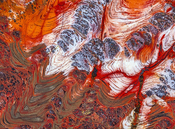
Biwabik Iron Formation stromatolites, made up of swirls of predominantly red and metallic hematite, bear witness to the previous presence of cyanobacteria 3.5 billion years ago. For at least half the time that life has existed on earth, bacteria were the only forms of life and, during this time, those with blue-green pigment that could photosynthesize (cyanobacteria) significantly changed the atmosphere by adding oxygen. Originally the earth's atmosphere was like that of Mars and Venus, with probably less than one percent oxygen. Photosynthetic cyanobacteria produced not only oxygen from carbon dioxide but also storable high-energy chemical compounds like adenosine triphosphate. These compounds permitted higher forms of life to evolve by allowing locomotion and other physiologic activities to develop. Courtesy: Norm Barker
We thank Professor Barker for taking his time in answering our interview questions. If you are interested in his images, please do check out some of his books which give detailed information on the images he took.